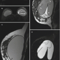
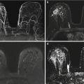
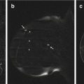
Fig. 4.1
Sagittal MRI images of the left breast. (a) T1-weighted fat-saturated image. (b) T2-weighted fat-saturated image. (c) T1-weighted fat-saturated post-contrast image, with corresponding computer-aided detection (CAD) kinetics analysis color overlay (d)

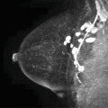
Fig. 4.2
Sagittal post-contrast subtraction maximum intensity projection (MIP) images of the left breast (a) and right breast (b)
4.2 Cowden Syndrome
Teaching Points
Cowden syndrome is an uncommon, autosomal dominant disease in patients with the PTEN gene mutation. It is characterized by multiple hamartomas of the skin, mucous membranes, brain, breast, thyroid, and gastrointestinal tract and increased susceptibility to breast cancer. The lifetime risk for breast carcinoma in patients with Cowden syndrome is estimated to be 25–50 %, compared with 12 % in the general female population.
Patients with Cowden syndrome can have bilateral multiple fibroadenomas, as illustrated in this case, which makes breast cancer detection challenging. Fibroadenomas usually enhance homogeneously, have associated T2 hyperintensity and can demonstrate nonenhancing septations. The degree of enhancement and T2 signal intensity can vary. The National Comprehensive Cancer Network (NCCN) recommends breast self-examination beginning at the age of 18 years and annual screening with breast MRI and mammography starting between 30 and 35 years of age and not earlier than 25 years. After establishing a baseline, any change should prompt a workup to exclude malignancy.
Image Findings

Fig. 4.3
MRI performed for multiple breast masses. A sagittal T1-weighted fat-saturated image (a) and a T2-weighted fat-saturated image (b) of the left breast demonstrate multiple masses with T1 hypointensity and T2 hyperintensity (arrowhead) and T1 hypointensity and T2 minimal to mild hyperintensity (arrows). A T1-weighted fat-saturated post-contrast image (c) shows homogeneous enhancement in these masses (arrows), with persistent delayed enhancement color-coded as blue on the CAD overlay (d)

Fig. 4.4
Sagittal post-contrast subtraction MIP images of the left breast (a) and right breast (b) show multiple masses in both breasts
4.3 History
4.4 Personal History of Breast Cancer
Teaching Points
Post-lumpectomy tumor recurrence rates are about 1–2 % per year, usually discovered later than 18–24 months after treatment. Detection of treatment failure in these women while still asymptomatic improves relative survival by 27–47 %.
The lumpectomy site is a common site of recurrence, as illustrated in this case. Patients with recurrence following initial treatment with lumpectomy and radiation are usually treated with salvage mastectomy. Breast MRI has a high sensitivity (94–100 %) in the detection of breast cancer. MRI screening of women with only a personal history of breast cancer was clinically valuable in finding malignancies in 12 % of the patients in a study published by Brennan et al. (2010), but personal history is generally not an accepted indication for screening breast MRI.
Image Findings

Fig. 4.6
Breast cancer recurrence. Sagittal T1-weighted pre-contrast (a) and T2-weighted (b) images of the right breast demonstrate post-radiation skin thickening (arrowheads) and the lumpectomy site (arrows), which is heterogeneously T2 hyperintense. (c) Sagittal T1-weighted FS post-contrast image with CAD color overlay demonstrates irregular thick rim enhancement with washout kinetics (red, arrows) and adjacent background parenchymal enhancement with persistent kinetics (blue)
4.5 History
33-year-old woman with BRCA1 mutation undergoing high-risk screening mammogram and breast MRI (Figs. 4.7, 4.8, 4.9, and 4.10).


Fig. 4.7
Craniocaudal (CC) mammographic view (a) and mediolateral oblique (MLO) mammographic view (b) of the left breast

Fig. 4.8
Sagittal MR images of the left breast. T1-weighted fat-saturated pre-contrast image (a) and post-contrast image (b) with CAD color overlay (c). (d) T2-weighted fat-saturated image
4.6 BRCA1 Mutation
Teaching Points
Teaching points: see next companion case.
Image Findings

Fig. 4.9
Screening mammograms. CC view (a) and MLO view (b) of the left breast demonstrate heterogeneously dense tissue. There is no suspicious mass, architectural distortion, or microcalcifications

Fig. 4.10
Screening breast MRI. The sagittal T1-weighted fat-saturated pre-contrast image (a) and post-contrast image (b) demonstrate a suspicious, enhancing irregular mass in the lower breast (arrow). CAD analysis (c) demonstrates washout enhancement kinetics. No mammographic correlate was noted. The corresponding T2-weighted fat-saturated image (d) demonstrates no T2-hyperintense correlate (arrow)
4.7 History
33-year-old BRCA2 mutation carrier undergoing screening breast MRI (Figs. 4.11 and 4.12).


Fig. 4.11
Sagittal MR images of the right breast. T1-weighted fat-saturated pre-contrast image (a) and post-contrast image (b), with corresponding CAD color overlay image (c). (d) T2-weighted fat-saturated image
4.8 BRCA2 Mutation
Teaching Points
Although there is no direct evidence that screening with MRI will reduce mortality, it is thought that early detection by using annual MRI surveillance, in addition to mammography, may be useful for high-risk women. The American Cancer Society recommends that screening MRI should be performed for women who are at high risk for breast cancer, based on certain factors:
Women with 20–25 % or greater lifetime risk of breast cancer, according to risk assessment tools that are based mainly on family history
Women with known BRCA1 or BRCA2 gene mutation
Women with a first-degree relative with a BRCA1 or BRCA2 gene mutation who have not had genetic testing themselves
Women who had radiation therapy to the chest between the ages of 10–30 years
Women who have Li-Fraumeni syndrome, Cowden syndrome, or Bannayan-Riley-Ruvalcaba syndrome or have first-degree relatives with one of these syndromes
BRCA1 and BRCA2 are tumor-suppressor genes that play a role in DNA repair; mutations in either gene are associated with an autosomal dominant inherited form of breast and/or ovarian cancer. The two cases illustrate high sensitivity of breast MRI in detecting clinically favorable early breast cancers in patients with high risk for breast cancer, including patients with BRCA1 and BRCA2 gene mutations. Lack of a hyperintense T2 correlate should elevate the level of suspicion, and biopsy should be performed in these high-risk patients even without a corresponding sonographic correlate.
Image Findings

Fig. 4.12
Mammographically and sonographically occult invasive ductal carcinoma (IDC). Sagittal T1-weighted fat-saturated pre-contrast image (a) and post-contrast image (b) and corresponding CAD color overlay image (c) demonstrate a homogeneously enhancing round mass (arrow) with washout kinetics. (d) No corresponding T2 signal hyperintensity was present. (Incidentally noted are metal artifact (arrowhead) and poor fat saturation (arrow) in the axilla.) The mass was not identified on second-look ultrasound. MRI-guided biopsy yielded IDC
4.9 History
33-year-old patient undergoing high-risk screening breast MRI (Figs. 4.13 and 4.14).


Fig. 4.13
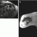
Axial T1-weighted fat-suppressed post-contrast image (a) and axial T2-weighted fat-suppressed image (b) of both breasts. Sagittal post-contrast subtraction image of the left breast (c) with CAD color overlay (d)

