1. Histology and Function
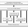
Bursae are cavities found or formed in areas subject to friction and lined by synovial membrane. The synovial membrane consists of two distinct layers: the inner intimal lining and the outer subintima. The intimal lining is 1-2 cells thick and contains synoviocytes, whereas the subintima is relatively acellular and consists of connective tissue containing scattered blood vessels, nerve fibres, fibroblasts, adipocytes, and a limited number of resident immune cells such as macrophages and mast cells.1,2 Two types of synoviocytes are identified in normal intimal lining.3 The type A synoviocyte is of macrophages lineage and derived from blood monocytes, while the type B synoviocyte is of fibroblast lineage and locally derived from subintimal fibroblasts.4,5 Type B synoviocytes have adapted to the production of a long-chain sugar polymer called hyaluronan and express several adhesion molecules, including vascular cell adhesion molecule-1 (VCAM-1), intercellular adhesion molecule-1, CD44 and β1 integrins.6 VCAM-1 expression may play a key role in cellular trafficking because its ligand is present on mononuclear leukocytes but not granulocytes, so the presence of VCAM-1 entrap macrophages and lymphocytes within the synovial membrane while allowing granulocytes (neutrophils, basophils, and eosinophils) to egress into the synovial fluid.7 The synovial membrane also allows diffusion of other elements of a plasma filtrate and components of its own to produce synovial fluid through contributions of proteoglycan, hyaluronan, and surface active phospholipids. Normal synovial fluid inhibits adhesion of synovial surfaces and reduces the coefficients of kinetic and static friction to a level ten times lower than the frictional coefficients of ice to ice. The hyaluronan is the main component responsible for the constant volume of bursal fluid during physiological loading because its rate of synthesis is dependent on the mechanical stress on type B synoviocytes.8 Hence, an increase in bursal fluid reduces mechanical stimulation on type B synoviocytes via synovial fluid cushion with a resultant reduction in the rate of synthesis of hyaluronan and vice versa. Unlike other serosal surfaces that also have non-adherent properties, synovial membrane is derived from ectoderm and does not contain a basement membrane due to the absence of ectatin in the intimal matrix.5
Related posts:

Stay updated, free articles. Join our Telegram channel

Full access? Get Clinical Tree


