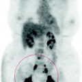
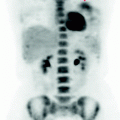
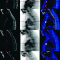
Fig. 39.1
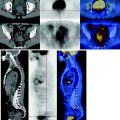
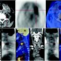
CT-PET: gross solid, inhomogeneous mass occupying the right anterior and medium mediastinum, with limited glucose metabolism
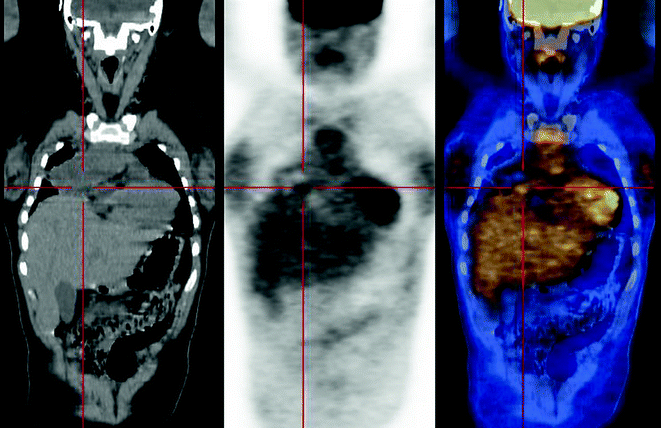
Fig. 39.2
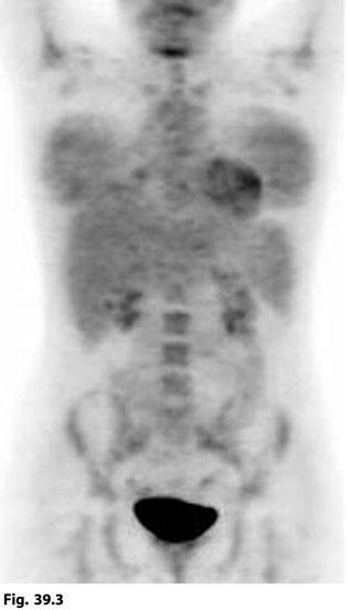
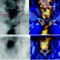
The coronal CT-PET image shows the solid mediastinal mass, with limited carbohydrate deposition (Fig. 39.2). In the MIP reconstruction focal lesions with a high uptake of glucose are not shown, in particular the mediastinal mass presents metabolic activity comparable to that of the liver (Fig. 39.3). Also observed widespread fixation of glucose in the bone marrow due to post-chemotherapy rebound. Clear increase in the metabolism of the mammary glands, typical of young women
