KEY FACTS
Imaging
- •
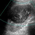
Thin-walled, distended tube; tube wall < 3 mm
- •
Convoluted or S-shaped, oval or pear-shaped, more dilated at fimbriated end
- •
Separate from uterus and ovaries
- •
Content anechoic; low-level echoes suggest acute pelvic inflammatory disease (PID)
- •
Thin endosalpingeal folds (~ 2-3 mm) protrude into lumen
- •
Incomplete septa: Short, linear echogenic projections into lumen from tubal kinking
- •
Waist sign: Indentation of opposing walls of dilated tubal structure resulting in appearance of waist
- •
Beads on string sign: Small hyperechoic mural nodules on transverse imaging
- •
Cogwheel sign: Thicker endosalpingeal folds in acute PID
- •
No color flow in endosalpingeal folds or wall
- •
Color flow in thickened tube suggests acute infection (as well as internal debris)
Top Differential Diagnoses
- •
Pyosalpinx (acute PID)/tuboovarian complex
- •
Cystic ovarian neoplasm
- •
Paraovarian cyst/peritoneal inclusion cyst
- •
Dilated bowel, acute appendicitis
Pathology
- •
Tubal obstruction from PID, endometriosis, appendicitis, or postpelvic surgery
Clinical Issues
- •
Usually asymptomatic, can present with pelvic or lower abdominal pain, severe pain may indicate adnexal torsion
- •
Must be distinguished from pyosalpinx or hematosalpinx based on tubal content and clinical picture
- •
Can result in infertility or ectopic pregnancy
Scanning Tips
- •
Cine clips are very useful for confirmation of tubular configuration
- ○
Look for incomplete septa and mural nodules in tubular, fluid-filled structure separate from uterus and ovaries
- ○
- •
3D-rendered US can help see tortuous structure, which is difficult to follow with 2D imaging
 , which appears as an incomplete septum on US. The fimbriated end
  is more dilated. Adhesions
  and hydrosalpinx are sequelae of pelvic inflammatory disease (PID).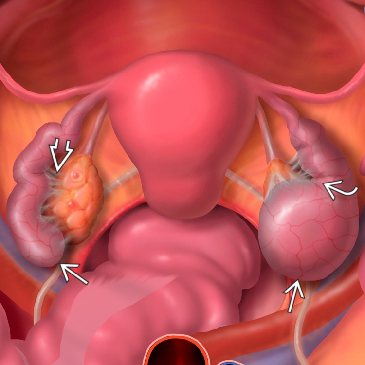
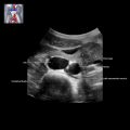
 with a dilated fallopian tube
  . The septa
  are the walls of the folded tube.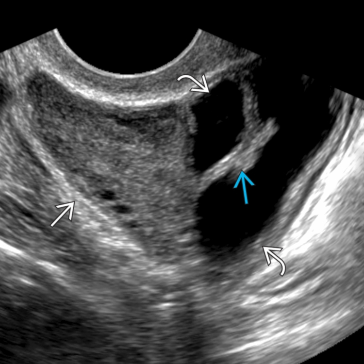
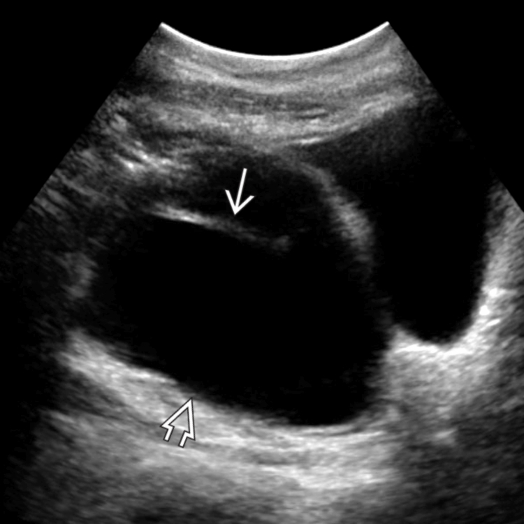
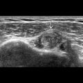
 with a thin, incomplete septum
  .
.
 . The uterus
  was normal. The incomplete septum
  is better seen as a kink in the tube.
Related posts:

Stay updated, free articles. Join our Telegram channel

Full access? Get Clinical Tree


