Hypertensive Basal Ganglia Hemorrhage
Kwaku A. Obeng
CLINICAL HISTORY
56-year-old male with a history of end-stage renal disease and hypertension, found unresponsive, and unable to move his right arm and leg.
FINDINGS
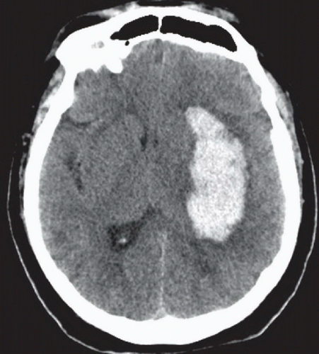
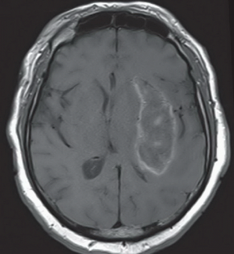
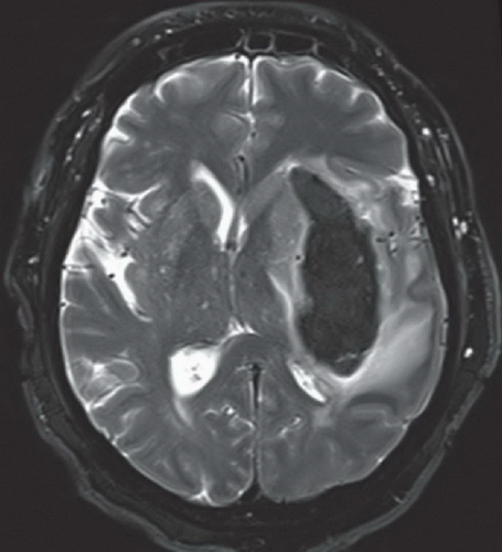
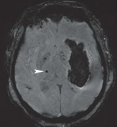
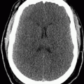
Figure 97A: Axial noncontrast CT image demonstrates a large, hyperdense hematoma centered in the lateral aspect of the left lentiform nucleus, compressing the left lateral ventricle and producing left to right midline shift. Figures 97B and 97C: Corresponding axial noncontrast T1W (Fig. 97B) and T2W (Fig. 97C) images demonstrate the hemorrhage to be predominately isointense to brain on T1WI and hypointense on T2WI centrally, indicative of deoxyhemoglobin. The periphery of the lesion demonstrates T1 shortening, indicating localized breakdown of hemoglobin into methemoglobin. The T2WI also demonstrates vasogenic edema around the hemorrhage. Though not shown, MRA did not demonstrate evidence of an aneurysm or arteriovenous malformation. Figure 97D: Axial SWI image demonstrates signal void throughout the hematoma. In addition, there is a small focus of signal dropout in the right thalamus (arrowhead), compatible with a remote microhemorrhage. Similar foci of signal dropout, suggestive of remote microhemorrhages, were also evident in the bilateral dentate nuclei (not shown).
Stay updated, free articles. Join our Telegram channel

Full access? Get Clinical Tree


