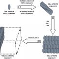
Fig. 2.1
Temperature ranges used for hyperthermia
The properties of the extracellular microenvironments of tumors are important in the outcome of hyperthermia treatments as will be discussed later. The unusual properties of the tumor environment compared to normal tissues are a product of rapid de novo cancer cell proliferation without adequate increase in angiogenesis or perfusion of the tissue area containing the tumor [9]. Tumor growth therefore outstrips the capacity of the blood supply, resulting in a build up of waste products such as metabolic acids and CO2, and depletion in O2 and glucose, as determined in the classical tumor pathophysiology studies of Pietro Gullino and co-workers [10]. The inadequacy of the tumor milieu is further exaggerated by a switch in tumor metabolism from oxidative phosphorylation to glycolysis (the Warburg Effect), an alteration that drastically increases glucose use and the production of acidic waste products [11, 12]. Growth of tumor populations away from the capillary networks also leads to tumor hypoxia, as O2 can only diffuse a short distance in tumors and is rapidly consumed by intervening cells [9]. The tumor microenvironment is also relatively immunosuppressive under unperturbed conditions. Tumor cell populations may evolve these properties through selection, by surviving and evading immunosurveillance during their origin and progression [13]. It is known that cancer cells can be essentially antigenic due to the presence within them of re-expressed embryonic antigens and the mutated proteins that seem a driving force in tumorogenesis [14, 15]. However, tumor cells appear in practice to develop a range of strategies to evade immune cell killing including “loss of self”, in which major histocompatability class I molecules are down-regulated making the cancer cell invisible to immunosurveillance. Further strategies to evade immunity include the secretion of the immunosuppressive cytokine interleukin-10 (IL-10), expression of Fas ligand on cancer cells to kill invading T cells and recruitment of immunosuppressive T regulatory cells (Treg) and myeloid suppressor cells (for review: [16]). Immunosuppression in the tumor microenvironment may also be a property of cancer stem cells, key tumor initiating subpopulations in cancer that are known to attract Treg cells, release IL-10 and show resistance to vaccines prepared from antigens expressed in the bulk tumor cell population [17].
2.2 Hyperthermia
2.2.1 Fever Range Heating (FRH)
The core body temperature of humans is normally maintained between 35.1 and 37.7 °C, although in the periphery of the body, including fingers and toes of humans and the paws of mice temperatures can be as low as 32 °C [18]. During fever , whole body temperature can rise as mentioned above, by 2–3 °C due to a cytokine-induced rise in the temperature set-point [2, 3]. These conditions can be reproduced artificially by whole body heating [5]. Experiments are usually carried out in mice at around 39.5 °C, a temperature well tolerated by healthy organisms [18, 19]. Direct cell killing is minimal at these temperatures and effects of FRH on tumor cells appear to be largely indirect. Hyperthermia in the FRH range does however cause profound modification of the activities of immune cells and may lead to an antitumor immune response [19]. Indeed FRH affects multiple immune cells, including the increased homing of dendritic cells to lymph nodes, increased cytokine production, elevated MHC class II levels, presentation of co-activating molecules to T lymphocytes, and proliferation of immune effector cells [20–23]. The molecular mechanisms involved in these changes appear to involve profound alterations in the mobility of molecules in the plasma membrane at FRH temperatures [19]. Of particular significance, FRH appears to allow molecules to enter lipid raft domains within the plasma membrane [19]. Lipid rafts are cholesterol and sphingolipid-rich plasma membrane microdomains that are known to contain a range of signaling molecules, including the innate immune stimulatory protein Toll-like receptor 4 [24]. Hyperthermia may thus exert its effects at FRH temperatures, at least partially by modifying membrane fluidity, modulating lipid raft behavior and clustering regulatory proteins to initiate cell signaling cascades that arise from the plasma membrane [19]. In addition, FRH alters tumor blood flow and may lead to increased passage of immune cells to tumors and the resolution of immunosuppressive tumor hypoxia [25]. It has been shown recently that FRH may play a profound role in immune killing of tumor cells by permitting cytotoxic lymphocytes to enter the tumor milieu after penetrating the tumor microvasculature [26, 27]. Tumor capillaries have been shown to pose a barrier to the entry of cytotoxic lymphocytes (CTL) into the tumor milieu and FRH appears to have the capacity to overcome this barrier through effects on interleukin-6 metabolism and extravasation of CTL in tumors [28]. It is tempting to speculate that fever may play an immunological adjuvant role in the responses of mammals to infection by pathogens and that use of FRH in cancer therapy may mimic a natural component of the innate immune response for stimulation of anti-tumor immunity. FRH used in a whole body context may be used in cancer treatment either alone or in combination with other modalities such as radiotherapy [5, 25].
2.2.2 Hyperthermia Range Heating
The “Hyperthermia Range” of heating is the one contemplated by exponents of this modality studied largely in the 1960s–1980s and comprises temperatures between 42 and 47 °C [1, 29] (Fig. 2.1). This temperature range appears to produce a set of cytotoxic lesions in cells that can defined using analysis by the Arrhenius equation as involving activation energies in the range required for protein denaturation [30]. Cell inactivation at these temperatures may involve direct killing due to increases in denatured and aggregated proteins in cells or indirect killing due to the triggering of programmed cell death mechanisms such as apoptosis by the proteotoxic stress [1, 31]. Killing in this range was found to be a regular, predictable process with an approximate doubling in killing for each degree rise in temperature . In addition there was found to be a profound difference in temperature sensitivity at 42 °C, compared to the higher end of the range and this appeared to be due to the ability of tumor cells to develop resistance (‘thermotolerance’) during heating at 42 °C but not at the higher temperatures [32, 33]. Resistance was later found to be due largely to the abundant expression of heat shock proteins (HSPs) , stress-inducible molecular chaperones that can refold protein aggregates generated in cells in hyperthermia and restore the quality of the proteome during treatment [34, 35]. In addition to playing intracellular roles in thermal protection as molecular chaperones , HSPs were subsequently found to be functional in the extracellular spaces and possess profound inflammatory and immunoregulatory properties [16].
Above and beyond the direct killing properties of heat discovered in vitro, it was found that conditions that might be expected to occur in the tumor milieu, hyperacidity and elevated lactate and CO2 powerfully profoundly sensitized cells to hyperthermia [4, 36]. Indeed hyperthermia at 42 °C and above led to a sharp decrease in tumor blood flow, conditions expected to exacerbate the poorly regulated tumor microenvironment [37]. Thus hyperthermia could be expected to be more effective in tumor cells in situ rather than in tumors in vivo. Indeed rates of apoptosis in prostate cancer cells after 43 °C hyperthermia vastly exceed rates observed under the same heating conditions in vitro [38, 39]. Thus the approach of using hyperthermia in this range with or without accompanying modalities remains highly promising in terms of favorable biological properties, although the clinical treatment of deep-seated tumors has proven technically very difficult and has been abandoned in many centers in the USA. Successful exceptions however exist and the utility of this heating range for clinical cancer has been proven [7, 8].
It could be predicted that hyperthermia in this range might be immunogenic due to release of the abundant levels of HSPs that accumulate in heating [40]. Hsp70 released during heating might contain tumor antigens and thus act like a molecular chaperone vaccine, transporting tumor antigens to antigen presenting cells and triggering activation of tumor-specific CTL [16, 41]. However, the effects of locally applied hyperthermia on tumor immunity are not consistent and both stimulation and inhibition of immunity are observed in this temperature range [42]. These findings may be related to our studies showing that, although apoptosis is a rare event in tumor cells exposed to hyperthermia in vitro by water bath heating, this mode of cell death appears to dominate, at least in prostate tumors heated in vivo [38, 39]. Apoptotic cell death is, almost by definition inhibitory to immunity and engulfment of apoptotic cells can lead to tolerance of the immune system to engulfed antigens [43]. The response of tumors to hyperthermia might thus involve competition between the immunogenic effects of Hsp70-peptide complexes and the tolerizing effects of apoptotic cells that occur in heated tumors in vivo. Significantly, the combination of hyperthermia in this range with immunotherapy leads to enhanced tumor cell killing in vivo and tumor regression . In melanoma, the combination of hyperthermia at 43 °C (using magnetite cationic liposomes) with intratumoral injection of DC led to DC activation and tumor regression [44]. Significantly heating in this mode was shown to lead to necrotic cell death . Similar results were seen in patients with advanced melanoma in which combined intratumoral injection of DC and hyperthermia led decreased tumor progression and immunostimulation [45]. Similar results were observed in prostate carcinoma [46]. Hyperthermia may modulate immunity by cell killing, altering HSP release from tumor cells and/or activating antigen presenting cells such as DC and altering cytokine release from tumor cells [20, 23, 40, 47]. In addition, hyperthermia has been shown to enhance the activities of molecular chaperone-based vaccines including high molecular weight heat shock proteins such as Hsp110 or Grp170 and lead to regression of tumors [48].
2.2.3 Ablation Range Hyperthermia
Thermal ablation of tumors such as hepatoma is carried out at temperatures exceeding 50 °C using radiofrequency (RF), microwaves and high intensity focused ultrasound [6, 49]. At 48–55 °C the mechanisms of cell killing appear to differ from the hyperthermia range and involve much lower activation energies for cell inactivation. These activation energies suggest a mode of cell death independent of protein denaturation and the authors ascribed them to effects on the integrity of DNA [50]. The precise molecular events involved in killing at these temperatures are not clear although it is known that the melting temperature of DNA in vitro is 87 °C, clearly well above the range studied by Landry and Marceau [1]. Other intracellular targets are also likely to be involved above 50 °C and we have observed a progressive increase in trypan blue uptake in PC-3 and LnCap prostate cancer cells from 45 °C, reaching uptake by 100 % of cells after 30 min at 50 °C [47, 51]. These findings indicate the loss of membrane integrity by 50 °C heating and our studies have shown that intracellular Hsp70 is passively released from the prostate cancer cells at this temperature [47]. Interestingly, 50 °Cappears to be the threshold temperature needed to trigger tissue coagulation and necrosis in ablation therapy [52]. Heating using the ablation approach may also be highly heterogeneous with, in the case of RF ablation, high temperatures that may approach 100 °C in tumor regions close to the RF antenna and significant cooler spots in the cancer [6]. Thermal ablation using this approach may thus subject tumors to each of the three hyperthermia ranges described above, depending on proximity of cancer cells to the heating antenna.
As absorption of necrotic cells by APC is thought to lead to immunity , one might predict that thermal ablation through its necrotizing effects on tumors would be immunogenic. Indeed treatment of hepatocellular carcinoma by RF ablation can activate tumor specific T cell responses [53, 54]. Hsp70 released from the necrotic cells may play a role in immunity under these circumstances [51]. Indeed, release of Hsp70 from cells undergoing necrosis has recently been shown to trigger specific antitumor immunity , activate a pro-inflammatory Th17 response, inhibit regulatory T cell responses and lead to tumor regression [55]. However, for eradication of secondary tumors by thermal ablation, combination with other immunogenic agents such as anti-CTLA-4 antibodies that block the effects of inhibitory T cell co-receptor CTLA-4 may be required [56, 57]. Indeed Waitz et al. [57] showed that inclusion of anti-CTLA-4 antibodies in the ablation protocol increased the ratio of effector T cells to inhibitory Treg cells in mouse TRAMP-C2 prostate cancers, circumstances that would favor a specific anti-tumor immune response.
2.3 Conclusions
Hyperthermia remains a form of therapy with great unexploited potential. Although the modality faces major technical challenges in uniform energy deposition and in being opposed by the powerful mechanisms of normal tissue temperature homeostasis, hyperthermia has major advantages arising from the tumor microenvironment . Some of the pathophysiological properties of the tumor milieu-low pH and glucose are known to strongly sensitize tumor cells to hyperthermia. The role of the antitumor immune response in hyperthermia is less simple. Exposure of tumor bearing animals to FRH, although not directly inducing cell death appears to be a powerful immune activator. However under conditions in which temperatures above 41 °C kill cells, the outcome in terms of tumor immunity is more complex. It would seem that when tumor cells undergo necrotic killing as observed at ablation-range temperatures, antitumor immunity is activated and tumor specific CTL are generated. Lower temperatures (below 50 °C) may lead to apoptotic killing, conditions likely to induce immune tolerance to the tumor cells. Combination of hyperthermia with immune adjuvants such as anti-CTLA-4 antibodies, dendritic cells, molecular chaperone-based vaccines or other activators may be recommended.
Related posts:
 In situ Tumor Ablation with Radiation Therapy: Its Effect on the Tumor Microenvironment and Anti-tumor Immunity
Radiofrequency Ablation in Cancer Therapy: Tuning in to in situ Tumor Vaccines
In situ Ablation of Solid Tumors by Electric Forces and Its Effect on the Tumor Microenvironment and Anti-tumor Immunity
Tumor-Localized Insult Delivered by Photodynamic Therapy and the Breakdown of Tumor Immunotolerance
In situ Tumor Ablation with Radiation Therapy: Its Effect on the Tumor Microenvironment and Anti-tumor Immunity
Radiofrequency Ablation in Cancer Therapy: Tuning in to in situ Tumor Vaccines
In situ Ablation of Solid Tumors by Electric Forces and Its Effect on the Tumor Microenvironment and Anti-tumor Immunity
Tumor-Localized Insult Delivered by Photodynamic Therapy and the Breakdown of Tumor Immunotolerance
 High Intensity Focused Ultrasound (HIFU) Ablation
 The Interrelationship Between Cryoablation, the Immune Response and the Tumor Microenvironment: Stimulatory and Suppressive Effects
Stay updated, free articles. Join our Telegram channel

Full access? Get Clinical Tree


