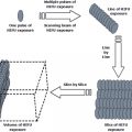
Fig. 7.1
Events and effector processes involved in host response to PDT of tumors
7.3.1 Onset and Advancement of Inflammatory Response
The early stage of inflammation in PDT-treated tumor tissue is focused on the lesion’s blood vessels. Its onset is hallmarked by the conversion of vascular endothelium from a nonthrombotic nonadhesive barrier to a proadhesive surface for inflammatory cells that becomes leaky for blood constituents [24]. Events securing this transformation include vasodilatation (consequent of increased production of arachidonic acid metabolites and nitric oxide synthase activation) that facilitates increased blood flow and leukocyte delivery, upregulated expression of leukocyte adhesion molecules, formation of gaps between endothelial cells, and platelet activation [18]. These changes set the stage for rapid invasion of neutrophils from circulation into the PDT-treated tumor [25, 26]. The appearance and extravasation of massive numbers of neutrophils is followed by the influx of activated mast cells and monocytes [25]. The main purpose of the engagement of these inflammatory cells is obviating the focal source of danger signals by disposing of the compromised tumor tissue elements and eliminating injured and dead cells as well as their debris. The most prominent visible clinical sign revealing the occurrence of inflammatory response is a pronounced tumor-localized edema manifested within several hours after PDT treatment.
Among the agents instigating and propagating the early stages of PDT-induced tumor inflammation are mediators liberated through the activation of the three major plasma cascade systems: complement, coagulation and fibrinolytic systems [17]. Other such rapidly generated agents independent of new gene expression that are participating in this phase of PDT response, are lysophospholipids and other lipid breakdown products of cellular membranes, arachidonic acid metabolites and histamine (the latter discharged from activated mast cells) [27]. They are in subsequent stages of post-PDT inflammatory process progressively replaced by mediators produced through upregulated gene expression resulting from triggered cellular signaling pathways; these include cytokines , chemokines and adhesion molecules [26,28,29].
7.3.2 Removal of Damaged Tumor Tissue
The key event of the acute inflammatory process in PDT-treated tumors is the removal of offending source i.e., the damaged tissue in these lesions. The inflamed mass that has to be disposed off consists of large numbers of dead and dying cancer and stromal cells (undergoing either apoptosis, necrosis, or autophagy) and their debris, as well as neutrophils and other inflammatory cells that have died after invading the site and performing their function. It is of essential importance that all these cellular corpses are promptly and efficiently cleared away because their continuous presence presents a serious threat, since it would perpetuate active inflammation , impair tissue healing and integrity, and could elicit autoimmune responses [30]. Due to its great physiological relevance, a process highly conserved through evolution has developed of regulated and efficient removal of dead cells that (particularly for apoptotic cells) has been termed efferocytosis (from Latin “effero”, meaning to carry to the grave) [31]. In situation when the host is suddenly faced with a large burden of dead cells in PDT-treated tumor, the professional phagocytes (macrophages and immature dendritic cells) that have high capacity of engulfment of dying corpses are indispensable in their role as efferocytes. At least some of PDT-associated DAMPs (lysophosphatidylcholine, S1P) are known to function as “find me” signals released from apoptotic cells that have a critical role serving not only as chemoattractants recruiting phagocytes to the site of cell death but also modulating their activation and differentiation and consequently influencing the ensuing inflammatory/immune response [32]. In addition to releasing soluble “find me” signals, dying cells in PDT-treated tumors become decorated with molecules exposed on their surface that are recognized by efferocytes as “eat me” signals [17, 33]. The latter include molecules normally localized inside the cells (phosphatidylserine, calreticulin, nucleic acids), altered existing molecules (changed glycosylation patterns, oxidized membrane lipids, and modified carbohydrates), and changed cell surface charges [31, 33, 34]. The recognition arm of the innate immune system can utilize a large number of engulfment receptors recognizing various “eat me” signals, and many of them appear to become engaged after PDT. There are two types of these receptors: (1) membrane-anchored on engulfing cells, and (2) soluble bridging molecules that opsonize dying cells to flag them as targets for efferocytes [34]. The efferocytosis of dying cells in PDT-treated tumors has a direct influence on subsequent host response progression, including not just the resolution of the inflammatory reaction but having also a critical impact on the development of adaptive immune response against PDT-treated tumor.
7.3.3 Resolution of Tumor Inflammation
Since prolonged inflammation increases the risk of inducing unnecessary damage to host tissue, a program ensuring its resolution is triggered without delay once the threat provoking the inflammatory response is resolved [35]. Key signals for downregulating inflammation are delivered by phagocytic macrophages, as their ingestion of dying cells in PDT-treated tumor prompts them to switch from producing pro-inflammatory cytokines to releasing mediators that suppress inflammation and engender the overexpression of factors that promote healing and survival/proliferation (to replace lost resident cells) [35, 36]. Among the most important of these anti-inflammatory agents are cytokines IL-10 and TGF-b, vascular endothelial growth factor (VEGF), cyclooxygenase-2 (COX-2), prostaglandins, matrix metalloproteinases and apoptosis inhibitor survivin [17, 37, 38]. Both IL-10 and TGF-b negatively regulate pro-inflammatory signaling by targeting transcription factor NF-kB and exert other multiple immunosuppressive effects [39, 40]. In addition, TGF-b promotes cell differentiation and growth, fosters matrix production, and stimulates fibrogenic remodeling essential in healing [41]. This activity it shares with the pro-angiogenic and tissue remodeling agents, VEGF, COX-2, prostaglandins, and matrix metalloproteinases, as well as pro-survival agents such as survivin [38, 42]. Unlike the phase of advancement of PDT-induced inflammatory response when vascular injury and inflammatory cell activity within PDT-targeted tumor tissue contribute to the eradication of cancer cells, the angiogenic and survival phenotype acquired in the inflammation resolution/healing phase may become permissive for tumor recurrence after PDT [17, 38]. Indeed, treatments targeting molecular responses that underline anti-inflammatory, pro-angiogenic or pro-survival activity can considerably improve the efficacy of PDT for tumor treatment, and indicate that postponing the resolution of tumor-destructive phase may be beneficial for therapeutic outcome. Some examples of applying this strategy include increased cure rates of PDT-treated tumors in mice injected with antibodies blocking IL-10 or TGF-b [17], COX-2 inhibitors [43,44], VEGF-blocking antibody [45], matrix metalloproteinase inhibitors [38, 46], and improved PDT response found upon interfering with survivin expression [42].
7.3.4 Host Response Effector Processes Supporting Inflammation
The inflammatory process focused on tumor tissue targeted by PDT is supported by at least several other host effector processes, including acute phase response , neural inflammatory reflex, and hormonal hypothalamic-pituitary-adrenal (HPA) axis (Fig. 7.1). All of them have overt systemic character, as they are mobilized to enable utilizing the resources from the entire organism, even those distant to the local PDT treatment site, to optimize the execution of host-protecting response [47]. The key feature of acute phase response is a striking increase or decrease in the release of a series of proteins called acute phase reactants due to altered biosynthetic profile in the liver or spleen and other sites [48]. Following PDT, a number of acute phase reactants are released and sequestered to the tumor site with the purpose to optimize and regulate the course of inflammatory events in the treated lesion [18, 47, 49]. A major group of these acute phase reactants are complement and pentraxin proteins specialized in opsonizing dying cells to facilitate their removal. Recently, heat shock protein 70 (Hsp70) was also recognized as acute phase reactant due to its upregulation in the liver and spleen of mice bearing PDT-treated tumors [50].
Glucocorticoid hormones and cytokine IL-6 were identified as key mediators involved in promotion of acute phase response induced by tumor PDT [47, 49]. Adrenal glucocorticoids appear to be released following the activation of HPA axis. It can be presumed that inflammatory cytokines released during PDT response are sensed as alarm signals by peripheral neural circuits operating neural inflammatory reflex [51, 52]. Such signals are propagated to the nucleus tractus solitarius in the brain stem by the vagus nerve, and then relayed to paraventricular nuclei in the hypothalamus activating the HPA axis. Release of serum corticosterone (primary glucocorticoid in mice) appears to peak at 1 h after PDT of tumors in these animals [47]. The up regulation of gene encoding glucocorticoid receptor (GR) and increased GR activity was demonstrated in PDT-treated tumors [47], which supports the concept that glucocorticoid influence the expression of multiple genes in these lesions. In such capacity they can have a dramatic effect on the activity of inflammatory and immune cells, have impact on apoptosis of tumor cells and influence the life span of mobilized immune cells [53]. This underscores the role of glucocorticoid hormones as vital controllers and regulators of the overall host response to tumor PDT.
7.3.5 Development of Adaptive Immune Response Against PDT-Treated Tumor
The above described PDT-elicited innate immune and inflammatory components of host response are antigen non-specific, although considerable numbers of tumor cells can be killed by invading inflammatory cells and activated complement system [18]. However, changes in tumor microenvironment associated with the execution of these host-protecting responses set the stage uniquely favorable for the development of adaptive (acquired) immune response specific for the antigens of PDT-treated tumor. It is also generally known that, in contrast to the same situation with normal tissue, inflammation of tumor tissue may diverge the course of host response towards a greater expression of immune rejection mechanisms; injured cancerous tissue (for instance cellular membrane structure) contains a greater abundance of potent danger signal molecules [54]. Key factors instigating adaptive immune response appear to be (1) the presence of overwhelming numbers of dead cancer cells within a very short time interval (overcoming the capacity of sequestered professional phagocytes to remove their corpses fast enough to avoid breaking immune tolerance), and (2) generation of photooxidative changes-modified antigenic fingerprint with tumor neoantigens exhibiting greater immunogenicity [55].
Burgeoning experimental evidence [6, 56] suggest that the tumor microenvironment changes following PDT stimulate phagocytes with engulfed dead cancer cells to transform (or mature) into antigen-presenting macrophages or dendritic cells , and migrate to tumor-draining lymph nodes. The antigens from the internalized tumor material bound to class II major histocompatibility complex molecules displayed on the membrane of these cells will there be recognized by T lymphocytes. Such activation of tumor-specific cytotoxic T cells (CD8+ T lymphocytes) followed by their clonal expansion will lead to the development of immunity against PDT treated tumor. Depending on tumor type, generation of these cytotoxic T cells and immune memory T cells may or may not involve active support of helper (CD4+) T cells , and their action may be augmented by natural killer cells [6, 57]. Thus PDT enables the host to overcome the immune ignorance of the targeted tumor and become capable of eliminating residual viable foci of cancer cells (persisting after initial response to PDT), which will impede the recurrence of the treated lesion. Moreover, disseminated cancer deposits distant to the area of PDT illumination may in some cases be eradicated by the induced immune response [58]. Thus, the capacity of eliciting antitumor immune response could have a critical role in the eventual outcome of PDT. Recently, experimental evidence was presented showing molecular identity of specific tumor antigens targeted by this immune response in mouse models [58, 59] and clinical setting [60].
Potentially one of the most significant achievements in the field of PDT is the development of cancer vaccines generated by this modality, which optimally exploits its capacity of induction of a potent antitumor immune response [55]. In contrast to standard PDT of tumors, in PDT vaccine protocols the patient is not administered the photosensitizing drug nor subjected to in situ tumor illumination; instead, a fragment of operatively resected tumor tissue is treated by PDT ex vivo producing the vaccine material that will be (after rendered safe by x-rays) injected into the patient. Such autologous whole-cell vaccines are optimally conditioned to target individualized, pertinent and even unique antigens in a patient-specific manner involving patient-matched MHC for recognition of tumor epitopes [55, 61].
7.3.6 Conclusion and Prospectives
Oxidative stress mediated by PDT causes a rapid and dramatic transformation in the microenvironment of treated solid tumors dominated by changes in blood and oxygen supply, and by elicited inflammatory response . Especially during the early period after PDT, the tumor microenvironment becomes more volatile undergoing acute changes influenced by numerous potent mediators engaged in the course of the elicited protective host response. Cure rates of PDT-treated tumors can be markedly improved by adjuvant agents targeting the expression of a variety of these mediators within the tumor microenvironment . The antigenic fingerprint of PDT-treated tumor enriched with photooxidative changes-induced neoantigens is captured in the corpses of cancer cells engulfed by activated phagocytes after the therapy. This information remains retained in the processed peptides as phagocytes are transformed into antigen-presenting cells , and is presented under favorable conditions to T lymphocytes enabling them to mount a vigorous adaptive immune response against PDT-treated tumor.
Related posts:
 Hyperthermia, the Tumor Microenvironment and Immunity
In situ Tumor Ablation with Radiation Therapy: Its Effect on the Tumor Microenvironment and Anti-tumor Immunity
Radiofrequency Ablation in Cancer Therapy: Tuning in to in situ Tumor Vaccines
In situ Ablation of Solid Tumors by Electric Forces and Its Effect on the Tumor Microenvironment and Anti-tumor Immunity
Hyperthermia, the Tumor Microenvironment and Immunity
In situ Tumor Ablation with Radiation Therapy: Its Effect on the Tumor Microenvironment and Anti-tumor Immunity
Radiofrequency Ablation in Cancer Therapy: Tuning in to in situ Tumor Vaccines
In situ Ablation of Solid Tumors by Electric Forces and Its Effect on the Tumor Microenvironment and Anti-tumor Immunity
 High Intensity Focused Ultrasound (HIFU) Ablation
 The Interrelationship Between Cryoablation, the Immune Response and the Tumor Microenvironment: Stimulatory and Suppressive Effects
Stay updated, free articles. Join our Telegram channel

Full access? Get Clinical Tree


