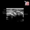
Hypoechoic Muscle Mass
ESSENTIAL INFORMATION
Key Differential Diagnosis Issues
Helpful Clues for Common Diagnoses
 Due to trauma, anticoagulation, or vascular malformation
 Anterior thigh is common location because it is susceptible to compression injury against femoral shaft and muscle contraction injury
 Initially hyperechoic or isoechoic
 Mild intramuscular hemorrhage may not be apparent on US, since ill-defined hyperechoic areas of blood blend with hyperechoic muscle
 Arises along course of peripheral nerve
 Well-defined, fusiform-shaped, hypoechoic mass
 Posterior acoustic enhancement (mild to moderate)
 Thickened entering or exiting nerve (very common)
 Split-fat sign due to separation of muscle fibers with enhanced visibility of fat at edge of tumor
 Mild to moderate hyperemia on color Doppler
 Cannot differentiate between schwannoma and neurofibroma based on US findings alone
 Try, however, to determine whether nerve is centrically or eccentrically placed relative to parent nerve
Helpful Clues for Less Common Diagnoses