CHAPTER 14 Ossicular Malformations
Epidemiology
Ossicular malformations are commonly associated with external auditory canal (EAC) malformations due to similar embryologic origins (for the auricle, first and second branchial arches; for the EAC, first branchial cleft; for the ossicular chain except stapes footplate, first and second branchial arches). Isolated ossicular malformations without outer ear abnormalities are rare but occur. Although autosomal dominant inheritance has been reported in some cases, these anomalies can be seen in association with such syndromes as Goldenhar and Treacher Collins.
Clinical Features
The outer ear anomalies that can be seen include microtia and atresia or stenosis of the EAC. In the presence of EAC abnormalities, there is an increased incidence of primary and secondary cholesteatomas. The ossicular malformations primarily produce a conductive type of hearing loss due to loss of sound transmission.
Pathology
Ossicular malformations have variable appearances on pathology that include hypoplasia, aplasia, or shortening of either a part of an ossicle or the entire ossicle itself. There can also be abnormal union between ossicles.
Treatment
Congenital conductive hearing loss due to ossicular deformities can be treated by either rehabilitation with a hearing aid or surgical reconstruction using a variety of ossicular prostheses.
Imaging Findings
CT
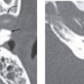
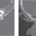
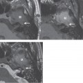
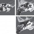
The ossicles most commonly malformed or absent include the stapes and incus. The stapes can reveal aplasia, hypoplasia, absence of head and crura, fusion of head to promontory, fixation of the footplate, and a columnar type deformity. Anomalies of the incus include aplasia, shortening, or malformation of the long process, absent or fibrous union of the incudostapedial joint, and fusion of the short process to the lateral semicircular canal. The malleus can be aplastic or have a deformed head. There can also be fusion at the incudomalleal joint or manubrium of the malleus, with the long process of the incus and the head of the stapes. Congenital fixation of the malleal head to the lateral epitympanic wall, called the malleus bar, is a rare anomaly.
Related posts:

Stay updated, free articles. Join our Telegram channel

Full access? Get Clinical Tree


