Spinal infections are challenging to diagnose and represent a life-threatening medical condition. Diagnosis is often delayed because of nonspecific accompanying symptoms. The role of interventional neuroradiology in spinal infection is double: diagnostic and therapeutic, consisting substantially of 2 main procedures, represented by spine biopsies and positioning of percutaneous drainage, which represent a minimally invasive, faster and more cost-effective alternative to open surgery procedures. This article will focus on the available state-of-the-art techniques to perform discovertebral image-guided biopsies in case of suspected infections and on image-guided placement of percutaneous drainage to treat infectious collections of the spine and paravertebral structures.
Key points
- •
The diagnosis of spinal infection is still today a challenging exercise; it can need one or more spine biopsies before the causative agent is isolated from specimens.
- •
To perform spine biopsies, there are several image-guidance available technologies, such as fluoroscopy, computed tomography, and MR imaging.
- •
Depending on the level of spine which is involved, a different approach and positioning of the patient should be used by the interventional radiologist.
- •
Antibiotic therapies eventually administered before the biopsy is performed can represent a main confounding factor; in those cases the sensitivity of the biopsy itself is further decreased.
- •
Percutaneous drainage of inflammatory collections eventually associated with spinal infections represents a safe and minimally invasive method to rapidly treat complications of spinal infections and can also be used to administer locally antibiotics.
Introduction
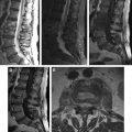
The diagnosis of spinal infections is still today a challenging exercise, and it can need one or more spine biopsies before the causative agent can be isolated from specimens. Imaging findings are rather characteristic, if compared to those associated with malignancy of the spine, but when facing cases with advanced pathology, it can be more difficult to assess the true nature of a process that can produce extensive disruption of spine components and their surrounding tissues.
Diagnosis can be difficult and is often delayed because of the nonspecific accompanying symptoms.
The role of interventional neuroradiology in spinal infection is double: diagnostic and therapeutic, consisting substantially of 2 main procedures, represented by spine biopsies and positioning of percutaneous drainage, which represent a minimally invasive, faster and more cost-effective alternative to open surgery procedures proposed for the same purposes.
This article will focus on the available techniques to perform discovertebral image-guided biopsies in case of suspected infections and on image-guided placement of percutaneous drainage to treat infectious collections of the spine and paravertebral structures.
Clinical cases from the personal experience will be reported, analyzing the imaging characteristics that lead one to formulate suspicion of spinal infection, with the interventional treatment adopted and follow-up evaluation.
A major focus will be applied on computed tomography (CT)-guided discovertebral biopsies, which represent a more diffuse way to perform safely and quickly spine biopsies in several pathologies of this anatomic district.
Introduction
The diagnosis of spinal infections is still today a challenging exercise, and it can need one or more spine biopsies before the causative agent can be isolated from specimens. Imaging findings are rather characteristic, if compared to those associated with malignancy of the spine, but when facing cases with advanced pathology, it can be more difficult to assess the true nature of a process that can produce extensive disruption of spine components and their surrounding tissues.
Diagnosis can be difficult and is often delayed because of the nonspecific accompanying symptoms.
The role of interventional neuroradiology in spinal infection is double: diagnostic and therapeutic, consisting substantially of 2 main procedures, represented by spine biopsies and positioning of percutaneous drainage, which represent a minimally invasive, faster and more cost-effective alternative to open surgery procedures proposed for the same purposes.
This article will focus on the available techniques to perform discovertebral image-guided biopsies in case of suspected infections and on image-guided placement of percutaneous drainage to treat infectious collections of the spine and paravertebral structures.
Clinical cases from the personal experience will be reported, analyzing the imaging characteristics that lead one to formulate suspicion of spinal infection, with the interventional treatment adopted and follow-up evaluation.
A major focus will be applied on computed tomography (CT)-guided discovertebral biopsies, which represent a more diffuse way to perform safely and quickly spine biopsies in several pathologies of this anatomic district.
Spine biopsies
Before proceeding with a biopsy of the spine, it is necessary to perform or review the radiological examinations that have raised its demand; these can be plain film radiographs, CT or MR imaging studies. The latter are usually preferred because of their higher sensitivity and specificity in detecting lesions of the spine and are often necessary to assess potentially associated spinal cord lesions of the involved spine tract.
The biopsy itself can be performed under several imaging guidances, such as fluoroscopy, CT and MR imaging; MR imaging-guided procedures are not yet widespread because of the costs and time spent to perform interventional procedures that, with the present technology, can be made more quickly and with superimposable safety under CT guidance, limiting significantly the radiation dose to which both patient and operator are exposed.
Fluoroscopy can represent an easy guidance method, but cannot show all the anatomic structures to be preserved during the procedure (ie, mainly nerve roots and vessels).
In case of multiple spine lesions, it is advisable to proceed to biopsy the level with fewer potential complications and with greater chances of easy and less traumatic percutaneous access.
Preparation
The patient must not drink or eat for a minimum of 8 hours prior to the procedure; it is necessary to acquire some important laboratory parameters to asses coagulation profile (prothrombin time [PT], partial thromboplastin time [PTT], international normalized ratio [INR]) and eventual allergies to drugs, especially local anesthetics and contrast media.
The procedure is often performed with a combination of local anesthetic (usually bupivacaine 1%) and intravenous conscious sedation using benzodiazepine. If a vertebral body has to be entered, the administration of local anesthetics around the periosteum is helpful to minimize the discomfort associated with the procedure.
Patient Positioning
Patient positioning depends on the level where the spinal biopsy is proposed; a prone position is often preferable for thoracic and lumbar lesions. In the case of a cervical lesion, a supine position is requested to gain anterolateral access to the cervical spinal tract; prone positioning should be considered safe to perform biopsy of the posterior cervical arches.
Depending on the patient clinical conditions, sometimes it is necessary to proceed to biopsy with an oblique prone or lateral decubitus.
Technique of Specimen Acquisition
There are 2 main possibilities that are almost always used together when performing a spine biopsy: aspiration biopsy and core biopsy.
Aspiration biopsy requires smaller caliber needles (18–22 G), while to perform core biopsy, greater cutting needles or bone biopsy needles are requested (10–14G); in the latter case, a small sample of tissue can be sent to pathologic examination, while the first method is more often preferred to produce specimens for microbiological analysis; actually, nothing prevents one from proceeding with aspiration through the same core biopsy needle used before.
These needles can be used with a tandem or coaxial technique.
Once the specimens have been acquired, to perform microbiological analysis, they must be placed in sterile containers and immediately sent to the laboratory; in case of aspiration biopsies it is of frequent observation the presence of blood, especially when the aspiration is performed after a core biopsy. It is important to consider that not only the frustules coming from the biopsy core site can be useful for microbiological or cytologic analysis, but also blood can be used for diagnostic purposes; thus it should not be considered as a waste and useless material. When multiple biopsy passes are performed and, if it is needed to rule out in differential diagnosis malignancy, part of the specimens can be put in containers with formalin 10% and sent to the pathology laboratory.
Tandem technique
This technique involves the use of 2 distinct needles; the first and smaller needle is used to administer local anesthetics alongside the trajectory planned for biopsy; once used, it is left in place to serve as a visual guide.
Coaxial technique
The localizing cannula is used to administrate local anesthetics and as a mechanical guide for the biopsy needle, which is coaxial to the cannula; using this technique, several passes with the biopsy needle are possible through the cannula, with major possibilities of diagnostic results and minor complication rates.
Percutaneous Approach to the Spine
The main determining factors to choose the way of percutaneous approach to the spine are represented by the lesion level and its dimensions.
The anterolateral approach is preferable only in the case of anterior cervical lesions; it requires great skills and experience as well as manual displacement of the neurovascular bundle of the neck. Otherwise, posterior access is safer in case of posterior cervical, thoracic, and lumbosacral lesions.
For thoracic biopsies, it is good practice to perform biopsies using a right lateral access, to avoid accidental injury to the aorta.
Posterior approach
Three ways of posterior approach can be distinguished ( Fig. 1 ).
Posterolateral
This approach is useful for lesions located within the vertebral body, intervertebral disc, or paraspinal tissues; it is clearly the most often used for biopsies in suspected spinal infections.
Transpedicular
This approach is used to access inside thoracic and lumbar vertebral bodies. The pedicle provides a safe way to access the vertebral body, but great care must be used to avoid its fracture, which can cause injury to the spinal cord or to the exiting spinal nerve root.
Trans-costo-vertebral
This approach is used only for lesions of the thoracic tract; it allows to one to reach the vertebral body, the intervertebral disc, and the paraspinal thoracic structures.
Postoperative Care
Immediately after the procedure, a sterile dressing is placed over the entry site(s). Patients must be observed for the next 2 to 4 hours, depending on the type of anesthesia used during the procedure, observing the site of access for eventual signs of active bleeding.
Diagnostic Efficacy
Literature show conflicting data about diagnostic reliability of spine biopsies in spinal infections, but it is a common consideration that spine biopsies, when made to diagnose spinal infections, have lesser diagnostic power than those performed in case of either primitive and secondary malignancy, and they are commonly reported to have positive results in a percentage varying between 15% to 70% of cases, depending also on the caliber of the needles used to perform such procedures. Actually, the most important factor influencing this result is surely represented by the fact that often patients with suspected spinal infections have already been under antibiotic therapy for several days. In such cases, it is possible to repeat the biopsy after at least 1 week without antibiotic therapy, and in those cases, literature data homogeneously show higher diagnostic power of the biopsies repeated after this measure.
Percutaneous drainage
When It Is Necessary?
Placement of a percutaneous drainage is requested to rapidly remove large fluid inflammatory collections that can be associated with advanced phases of spinal infections, to reduce local inflammation, and accelerate healing of the infection itself; they can be used also to introduce directly in situ antibiotics and produce materials for microbiological testing.
Technique
Once the level of the collection and its anatomic relationships with the nearest structures to be preserved have been identified, the drainage is placed using the same tricks and techniques used for biopsy and exposed earlier; it is almost always used a posterolateral approach to reach the collection, which can affect the psoas muscles and/or other minor paravertebral muscles.
The same precautions taken for the execution of spine biopsies must be taken in account for placement of a drainage (ie, evaluation of coagulation parameters and allergies).
6–8 F drainage catheters can be used, and they are more safely placed using CT guidance. Great care has to be used to avoid the nearest structures that could be damaged while inserting the drainage catheter. Once positioned, the catheter can be connected to an airtight bag or a container with internal negative pressure so as to ensure continuous aspiration.
Once the collection is resolved and the antibiotics have been injected locally, the drainage can be safely removed.
Related posts:


Stay updated, free articles. Join our Telegram channel

Full access? Get Clinical Tree


