9 Image Resolution: Pixel and Voxel Size
A pixel represents the smallest sampled 2D element in an image. It has dimensions given along two axes in millimeters, dictating in-plane spatial resolution. Pixel sizes range in clinical MRI from millimeters (e.g., 1×1 mm2) to submillimeters. A voxel is the volume element, defined in 3D space. Its dimensions are given by the pixel, together with the thickness of the slice (the measurement along the third axis). Slice thicknesses in clinical MRI vary from a maximum near 5 mm, achieved using 2D multislice imaging, to submillimeter, achieved with 3D scan techniques.
MRI spatial resolution, which determines the radiologist’s ability to distinguish structures as separate and distinct from each other (together with image contrast), is inherently related to the acquired voxel volume. In the simplest case, the field of view (FOV), acquisition matrix, and the slice thickness determine voxel volume. The pixel size (FOV/matrix) determines the in-plane resolution. Reducing the FOV, increasing the matrix number, or reducing the slice thickness results in an image with reduced voxel volume. SNR is directly proportional to voxel size (assuming that the number of phase-encoding steps is held constant). Small voxels produce MR images with high spatial resolution but lower signal-to-noise ratio (SNR) and thus may appear “grainy” compared with images acquired with a larger voxel volume.
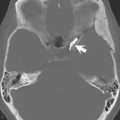
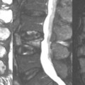
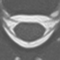
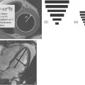
The images shown in Fig. 9.1
Related posts:

Stay updated, free articles. Join our Telegram channel

Full access? Get Clinical Tree


