Study
Type
No. of patients
PET
Conventional imaging
Sensitivity (%)
Specificity (%)
PPV (%)
NPV (%)
Accuracy (%)
Sensitivity (%)
Specificity (%)
PPV (%)
NPV (%)
Accuracy (%)
Lee et al. [10]
P
99
84
70
93
48
82
90
71
94
60
87
Rodriguez et al. [11]
P
16
80
82
67
90
–
–
–
–
–
–
Petrowsky et al. [12]
P
61
100
33
90
6
53
71
33
90
7
56
Yamada et al. [13]
R
14
69
–
90
–
64
–
–
–
–
–
In addition to the importance of accurately localizing the primary tumor, the detection of regional and distant metastasis is crucial to the determination of initial treatment strategies. Lymph node involvement and presence of metastatic lesions may preclude surgical resection. With respect to the detection of regional lymph nodes, PET/CT does not seem to offer a significant advantage over conventional imaging with reported sensitivities ranging from 12–82 to 24–80 % for PET/CT and conventional imaging, respectively [10, 12] (see Table 2). There is a clear and significant advantage, however, of PET/CT for the detection of suspected and unsuspected distant lesions, with reported sensitivities ranging from 95–100 to 25–63 % for PET/CT and conventional imaging, respectively [10, 12] (see Table 3).
Table 2
Detection of nodal metastasis in gallbladder and cholangiocarcinoma by PET/CT: comparison with conventional imaging
Study | Type | No. of patients | PET | Conventional imaging | ||||||||
|---|---|---|---|---|---|---|---|---|---|---|---|---|
Sensitivity (%) | Specificity (%) | PPV (%) | NPV (%) | Accuracy (%) | Sensitivity (%) | Specificity (%) | PPV (%) | NPV (%) | Accuracy (%) | |||
Lee et al. [10] | P | 99 | 82 | 95 | 94 | 85 | 89 | 80 | 79 | 78 | 81 | 79 |
Petrowsky et al. [12] | P | 61 | 12 | 96 | 67 | 64 | 64 | 24 | 86 | 50 | 65 | 62 |
Table 3
Detection of distant metastasis in gallbladder and cholangiocarcinoma by PET/CT: comparison with conventional imaging in prospective studies
Study | Type | No. of patients | PET | Conventional imaging | ||||||||
|---|---|---|---|---|---|---|---|---|---|---|---|---|
Sensitivity (%) | Specificity (%) | PPV (%) | NPV (%) | Accuracy (%) | Sensitivity (%) | Specificity (%) | PPV (%) | NPV (%) | Accuracy (%) | |||
Lee et al. [10] | P | 99 | 95 | 95 | 86 | 98 | 95 | 63 | 94 | 75 | 89 | 87 |
Petrowsky et al. [12] | P | 61 | 100 | 100 | 100 | 100 | 100 | 25 | 100 | 100 | 85 | 85 |
As a result of staging by PET/CT, treatment plans that were based on previous conventional staging were modified in up to 17 % of patients, whereby the detection of unsuspected metastasis by PET/CT led to non-surgical treatment, sparing unnecessary resection in patients deemed resectable following conventional work-up [10, 12] (see Figs. 1, 2, 3).
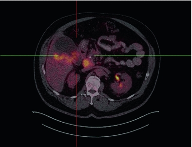
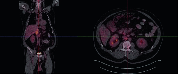
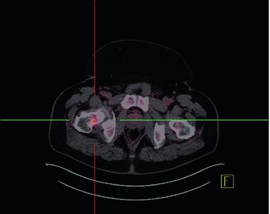
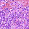
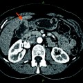
Fig. 1
54 year-old male with a newly found gallbladder mass. Axial fused staging 18F-FDG PET/CT image demonstrates intensely FDG-avid gallbladder wall thickening involving the posterior wall (SUVmax 10.1) and the hepatic parenchyma
Fig. 2
Coronal (left) and axial (right) fused PET/CT images demonstrate FDG-avid lymphadenopathy involving the aortocaval nodes with the highest SUVmax of 6.6
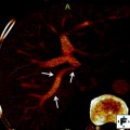
Fig. 3
Intense focal FDG uptake is noted in a lucent lesion involving the right femoral neck and is highly suspicious for malignant involvement with a SUVmax of 7.2
Much more research is warranted before definitive conclusions can be made; however, although limited, the data depict superiority of PET/CT for the staging of gallbladder cancer, especially in cases of equivocal conventional imaging, for evaluating metastatic disease, and for deciding on the initial treatment strategy.
4.2 Restaging
Very little data are available in the restaging setting, as such; the utility of PET/CT cannot be evaluated to a great extent. One prospective study compared PET/CT with MDCT for evaluating residual disease in patients with incidental gallbladder cancer [14]. Twenty-four patients with incidental gallbladder cancer who were suitable for surgery were recruited for the study [14]. For detecting residual disease, the authors reported a sensitivity and positive predictive value (PPV) of 28.5 and 20 % for PET/CT and 42.8 % each for MDCT, respectively [14]. Despite the low values, PET/CT was able to detect occult metastatic and loco-regional disease missed on MDCT, emphasizing the importance of using multimodality imaging in a complementary fashion as concluded by the authors as well [14].
5 Utilizing 18F-FDG PET/CT for Initial Treatment Strategy in Extra-Hepatic Cholangiocarcinoma
5.1 Staging
Similar to gallbladder cancer, malignancies of the biliary tract are also rare and have poor prognosis. There have been more studies, however, evaluating the role of PET/CT in biliary tract cancer, with the greatest advantages reported to be in detecting metastatic disease and selecting candidates for surgery [15]. Preoperative imaging is, thus, an integral part of initial assessment.
With respect to staging of the primary tumor, PET/CT seems to have limited or no clear advantage over conventional imaging techniques such as CT and MRI. This may be due to the overlap in FDG uptake between biliary tract malignancies and benign inflammatory lesions, especially in patients with primary sclerosing cholangitis (see Table 4).
Table 4
Detection of primary cholangiocarcinoma tumor by 18F-FDG PET/CT
Study | Type | No. of patients | Sensitivity (%) | Specificity (%) | PPV (%) | NPV (%) | Accuracy (%) |
|---|---|---|---|---|---|---|---|
Alkhawaldeh et al. [16] | R | 65 | 94 | 83 | 94 | 83 | 91 |
Corvera et al. [17] | P | 93 | 69 | 67 | – | – | – |
Kim et al. [18] | P | 123 | 81 | 79 | 95 | 44 | 81 |
Li et al. [19] | P | 17 | 59 | – | 100 |