10 Imaging Basics: Signal-to-Noise Ratio (SNR)
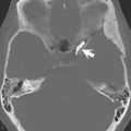
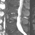
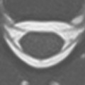
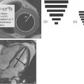
The images in this case illustrate the critical concept of signal-to-noise ratio (SNR) in MR SNR, as the term implies, is the ratio of MR signal to noise, specifically for the spatially encoded voxel. MR images acquired with a low SNR appear somewhat “grainy” to the eye (Fig. 10.1A and Fig. 10.2A) especially when compared with images acquired with higher SNR (Fig. 10.1B and Fig. 10.2B). However, what is most relevant clinically is the contrast-to-noise ratio (CNR), not the SNR The higher the contrast between two structures, the less SNR required for differentiation. To distinguish tissues that have similar contrast, high SNR is required. For example, the difference between gray and white matter (small white arrows, Fig. 10.2B) is much easier to see in Fig. 10.2B (with higher SNR) than in Fig. 10.2A.
What determines the signal in a spatially encoded voxel? MR signal is directly proportional to the size of the voxel. The larger the voxel, the greater the number of hydrogen protons and thus the greater the MR signal. Larger voxels, however, result in reduced spatial resolution (see Case 9
Related posts:

Stay updated, free articles. Join our Telegram channel

Full access? Get Clinical Tree


