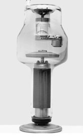
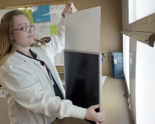
CHAPTER 8 On completion of this chapter, you should be able to: • Describe the x-ray tube and name its two main components. • Explain the basic process of x-ray production. • Explain the two general types of digital imaging systems. • Describe screen-film systems. • Describe the function of the fluoroscope. • Describe the function of computed tomography (CT). • Explain how an image is made in nuclear medicine. • Describe the use of portable radiographic and fluoroscopic units. • Discuss how an image is formed in sonography. • List the two types of information obtained by using magnetic resonance imaging (MRI). • Explain the primary use of positron-emission computed tomography (PET). X-rays are not stored, nor do they come from radioactive materials. The radiographer manufactures x-rays for each exposure using technical factors manipulated on the x-ray control panel. X-rays are produced by a series of energy conversions. The primary items needed for the production of x-rays are: (1) a source of electrons, (2) a means to accelerate the electrons, and (3) a way to bring the electrons to a sudden stop. All of these items are accomplished in the x-ray tube. (Details concerning the process of imaging are presented in Chapter 10.) The x-ray tube is an evacuated glass bulb with positive (anode) and negative (cathode) electrodes (Fig. 8-1). The anode is an electrode toward which negatively charged electrons migrate. The cathode is a filament that gives off electrons when heated (i.e., the source of electrons). As several thousand volts of electricity are applied to the tube, these electrons are driven across a short distance at a very high speed (i.e., the means to accelerate the electrons) and strike the anode with high kinetic energy (i.e., the way to bring the electrons to a sudden stop). Because energy can be neither created nor destroyed, an energy conversion takes place; this energy conversion is the result of the sudden deceleration of the electrons at the anode. Heat is the primary by-product (>99%) of this energy conversion. However, x-rays are also produced (<1%), and they emanate from the tube in all directions. The x-rays exit the tube housing through a device consisting of open lead shutters called a collimator. In computed radiography (CR), the x-rays exit the patient and strike a cassette containing an imaging place (IP) (Fig. 8-2). The IP is coated with a substance known as a photostimulable phosphor. This phosphor becomes excited as a result of the deposit of x-ray energy, and it remains so until the cassette is placed in a reader. Once inside the reader, the IP is scanned with a laser beam, releasing the x-ray energy that is then converted to a visible image by the computer. The resulting image is viewed on a high-resolution monitor.
Imaging Equipment
X-ray tube
Digital imaging
![]()
Stay updated, free articles. Join our Telegram channel

Full access? Get Clinical Tree


Radiology Key
Fastest Radiology Insight Engine