Child abuse and neglect is a serious clinical and socioeconomic problem that is sometimes underestimated. One of the most devastating forms is abusive head trauma. This review addresses the radiological workup in cases of suspected child abuse. The use of all modalities, and their advantages and disadvantages, is discussed. A special section is devoted to the radiological report in cases of child abuse, as a clinical record and a legal document.
Child abuse is defined by the World Health Organization as “Child abuse, sometimes referred to as child abuse and neglect, includes all forms of physical and emotional ill-treatment, sexual abuse, neglect, and exploitation that results in actual or potential harm to the child’s health, development or dignity. Within this broad definition, 5 subtypes can be distinguished – physical abuse; sexual abuse; neglect and negligent treatment; emotional abuse; and exploitation.” Although radiologists can potentially be confronted with all 5 forms of child abuse and neglect (CAN), this article deals exclusively with physical abuse.
The exact incidence and prevalence of CAN are unknown, because researchers use their own definitions of such abuse, which makes comparison between studies difficult. Among such studies are the National Incidence Study of Child Abuse and Neglect in the United States, the Canadian Incidence Study of Reported CAN, and the Dutch Prevalence Study of Maltreatment of Youth. In these 3 studies the incidence was reported to range from 17.2 to 30.0 per 1000 children. Another way of looking at the magnitude of this problem is presented by Lord Laming, who in his report on the tragic death of Victoria Climbié, stated “I have no difficulty in accepting the proposition that this problem (deliberate harm to children) is greater than that of what are generally recognized as common health problems in children, such as diabetes or asthma.”
Besides an immediate impact on the child’s health status, which can be severe or even lethal, there are serious social and medical long-term consequences of CAN. In the Adverse Childhood Experiences study, Felitti and colleagues found a graded relationship between the number of categories of adverse childhood exposures and adult health risk behaviors and diseases. Although beyond the scope of this publication it is of general interest to address the potential economic cost of CAN. In a 2010 publication Corso and Fertig presented corrected data for the United States, reporting that the annual cost, both direct and indirect, of CAN is a staggering $65,139,889,962. This figure does not even take into account the costs associated with mortality or reduced life expectancy.
In evaluating child abuse, a multidisciplinary approach is essential. Ideally, a team, which may be called the child advocacy team (CAT), consisting of all involved specialties including pediatric radiology should be involved. The CAT should support the attending physician and, if necessary, ask for help from a pediatric forensic physician. It interesting to note that the Royal College of Pathologists (RCP), at a meeting specifically aimed at the discussion on the cause of abusive head trauma (AHT), agreed that “in the current state of knowledge the presence of ‘the triad’ [see later discussion], even in its ‘characteristic’ form, should not be regarded as absolute proof of traumatic head injury in the absence of any other corroborative evidence.” The evaluation of child abuse is often challenging; the combined opinion of many specialists in the CAT lends credibility and certainty to the diagnosis.
Because this edition of Radiological Clinics of North America is devoted to imaging of the neonate, we focus on the most prevalent finding in physical abuse at the age: AHT. Given the importance of secondary findings in AHT, imaging of fractures is also discussed. Although the neonatal period is only the first month of life, most of the material discussed in this article can also be applied to the infant.
AHT
AHT is one of the most devastating forms of child abuse, and in children less than 2 years it is the leading cause of death in CAN. In 1946, the pediatric radiologist, J. Caffey reported on the unusual association of long bone fractures and subdural hematomas (SDHs) and suggested these 2 injuries may be secondary to child abuse, a radical idea at the time. Later, in 1962, Silverman, a radiologist, and Kempe, a pediatrician, and others described the battered child syndrome in a study that described the results of direct trauma on the child, renewing general interest in the problem of child abuse. In 1972, the shaken baby syndrome term was introduced in another study by J. Caffey. In his seminal paper, the classic triad consisting of encephalopathy, subdural hemorrhages, and retinal hemorrhages was first presented. However, the term shaken baby syndrome is not just a description of radiological findings but also implies an underlying mechanism (ie, shaking). In recent years, there has been much debate about the cause of AHT; in particular, whether shaking alone could lead to the findings seen on imaging. It was therefore deemed inappropriate to continue the use of shaken baby syndrome, and in 2009, the American Academy of Pediatrics (AAP) proposed the use of AHT instead. Other terms that are used besides AHT are nonaccidental head injury and inflicted traumatic brain injury.
In Great Britain the Crown Prosecution Service issued this statement on the diagnosis of AHT: “Each case will clearly turn on its own facts but it would appear unlikely that a charge of murder can be justified where the only evidence available is the triad of injuries” (the triad of injuries referred to is the so-called classic triad in AHT). Furthermore, it specifically states that “Cases of alleged non-accidental head injury are fact specific and will be determined on their individual facts. All the circumstances, including the clinical picture, must be taken into account.” This statement means, as stated in the introduction, that the diagnosis of AHT should be made only in close collaboration with a CAT. In each case of suspected AHT a complete clinical history and thorough physical examination are mandatory. Mcguire and colleagues performed a systematic literature analysis in which 8151 publications were identified and 14 of 320 reviewed publications were included. In total the study population consisted of 1655 children with neurotrauma (799 with AHT). From their data these investigators concluded that apnea (positive predictive value [PPV] 93%, odds ratio [OR] 17.06, P <.001), retinal hemorrhages (PPV 71%, OR 3.504, P = .03) and rib fractures (PPV 73%, OR 3.03, P = .13) were significant positive predictors for AHT. Although the presence of retinal hemorrhages is strongly related to AHT, the discussion of this topic falls outside the scope of this article and the interested reader is therefore referred to relevant publications.
Clinical presentation
The clinical presentation of children with AHT can be variable, rendering it difficult to make an accurate diagnosis in many cases. Jenny and colleagues have shown in a series of 173 abused children with head injuries, 54 (31.2%) cases were initially misdiagnosed. More important than these statistical findings is these investigators’ finding that 4 of 5 deaths might have been prevented by a timely recognition of CAN. This finding is in keeping with other publications on the diagnosis of CAN.
According to Minns and Busuttil, 4 main presenting patterns of AHT are seen:
- 1.
Hyperacute encephalopathy:
Approximately 6% of all children present with hyperacute encephalopathy, otherwise known as a cervicomedullary syndrome. These are mostly young children with acute brainstem conditions (ie, apnea and cerebral edema). In many, if not most cases, these children are dead on arrival in the hospital.
- 2.
Acute encephalopathy:
Most AHT cases, approximately 53%, are cases of acute encephalopathy. At presentation, these children have a low level of consciousness, increased cranial pressure, convulsions, apnea, hypotonia, anemia, or shock. Often additional injuries, such as rib fractures, are found. This pattern of AHT was originally described by J. Caffey as the shaken baby syndrome.
- 3.
Subacute nonencephalopathy:
This group of patients, approximately 19% of all cases, present with similar but less severe symptoms as seen in acute encephalopathy.
- 4.
Chronic extracerebral presentation:
Approximately 22% of all patients with AHT present with growing head circumference and mild symptoms of raised intracranial pressure (eg, failure to thrive and behavioral problems).
Imaging
Conventional Radiography
Historically, conventional radiographs of the skull were an important diagnostic tool. However, with the advent of computed tomography (CT), the use of skull radiographs in the trauma setting has been abolished. Several studies have shown that the sensitivity of skull radiographs for intracranial conditions is poor. The Children’s Head injury Algorithm for the prediction of Important Clinical Events (CHALICE) study, a prospective diagnostic cohort study of 22,772 children, assessed the value of the conventional skull radiograph. In this study, a total of 5318 skull radiographs were obtained, and in 259 cases the radiological report stated the presence of a fracture. Of these fractures, 44 (17%) were missed by emergency physicians. In 59 cases (1% of normal radiographs) a fracture was diagnosed by emergency physicians but read as normal by the radiologist. A subpopulation of 98 children with abnormalities (not otherwise specified) on CT also had a skull radiograph. In these patients, radiography had a sensitivity of 77% (95% confidence interval [CI] 67%–85%, 75 fractures in 98 patients) for a positive condition on CT. In a series of 47 children who sustained AHT, studied by Rao and colleagues, 31 (65.9%) did not have a skull fracture. In a study by Merten and colleagues among children with AHT, a skull fracture was seen in 45% of cases and intracranial conditions were seen in 56% of children with a skull fracture. Based on these and other publications, it can be concluded that the continued use of skull radiographs for children with acute head injury is not warranted. This finding is in keeping with previous studies. The AAP states in their position paper on the management of minor closed head injury that the skull radiograph has only a limited role in the evaluation of these children.
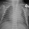
In child abuse, skeletal radiography is used to show the presence of occult trauma and clinical evident conditions and aid in the diagnosis of underlying disease ( Fig. 1 ). In rare cases, a skull fracture in the plane of scanning can be missed on CT. In these cases, the fracture may be visible on the skull radiographs. The reporting radiologist should be aware of sutural anatomy (ie, not only the normally seen sutures but also the normal variants, because these can resemble fractures). In a significant proportion of cases of AHT, skull fractures are seen.
Most skull fractures, whether from accidental trauma or CAN, are linear fractures; this finding is not discriminating. It has been reported that when there are bilateral fractures, multiple fractures with depression and diastases greater than 3 mm, depression fractures, fractures with diastases of the fracture lines, or occipital fractures, child abuse should be considered in the differential diagnosis ( Fig. 2 A and B). No single skull fracture is pathognomonic for child abuse.
In the most recent guidelines of the American College of Radiology (ACR) and the joint guideline from the Royal College of Paediatrics and Child Health (RCPCH) and Royal College of Radiologists (RCR), the skull radiograph is a mandatory part of the skeletal survey. This statement implies that even when a CT of the head has been performed, a skull radiograph should be obtained in all cases.
Ultrasonography
Cranial ultrasonography, which is widely adopted by pediatric radiologists because of its clear benefit to patient care, has no role in the primary detection of child abuse. Because of the convexity of the skull, SDH can be missed and detection of subarachnoid hemorrhage (SAH) is virtually impossible.
However, there are instances when the pediatric radiologist is asked to perform a cranial ultrasonographic study. This request may be in a situation when the patient is unstable and an urgent bedside examination is necessary. Change in status such as enlarging head circumference, at which time an SDH may be seen, may be the indication for the ultrasonographic examination. Using color Doppler imaging it is possible to discern between an SDH and benign enlargement of the subarachnoid space, a common finding in children who are referred for large cranial circumference ( Fig. 3 and Fig. 4 A, B). Jaspan and colleagues presented 6 children in whom contusional tears of subcortical white matter were detected using high-resolution ultrasonography, a finding that indicates AHT in these investigators’ opinion. Another use of cranial ultrasound, using a high-resolution probe, is for follow-up of SDH. When properly used this technique can obviate repeat CT or magnetic resonance (MR) imaging scanning.
CT
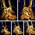
CT is the first-line imaging tool in the setting of traumatic brain injury, irrespective of the cause of trauma. With its wide availability and short scan times, it can readily identify acute conditions needing neurosurgical intervention. Images should be acquired using thin collimation, necessary for three-dimensional (3D) reconstruction. The images should be reconstructed with a maximum slice thickness of 5 mm in both a soft tissue algorithm (with, eg, window width (WW)/window level (WL) 80/25) and a bone algorithm (with, eg, WW/WL 3000/300). Standard 3D reconstructions, which should be obtained in all cases of AHT, are useful for showing radiological findings, especially (but not exclusively) to nonmedical personnel ( Fig. 5 ). The use of more advanced 3D techniques has been described in the literature but has not been validated in large samples.
Noncontrast-enhanced CT has good sensitivity for acute hemorrhage and midline shift ( Figs. 6 and 7 ). However, it is less sensitive, especially in the acute stage, for nonhemorrhagic intracranial conditions such as shear injury and perfusion disorders. In adults, the use of perfusion CT has been described in the evaluation of mild head injury. Metting and colleagues reported that disturbed cerebral perfusion, in patients with normal noncontrast-enhanced CT scans, was related to the severity of injury and outcome. This condition has not been studied in children and, given the lack of evidence, perfusion CT should not be performed in children with neurotrauma. In general, there is no indication for contrast-enhanced CT scanning. If a vascular lesion is considered, MR imaging angiography should be the modality of choice.
Subdural hemorrhage is seen in a significant proportion of children with AHT. Subdural hemorrhage is found in up to 90% of autopsied cases of AHT. In most instances it consists of a thin film of blood, over the cerebral convexities, often bilaterally. In young children, unlike adults, the subdural hemorrhage often extends into the posterior interhemispheric fissure ( Fig. 8 ). On a CT scan, a small amount of interhemispheric subdural hemorrhage can be visualized that might be missed at autopsy because of its location. However, compared with autopsy, CT is less sensitive in detecting small convexity subdural hemorrhages, a result of both beam hardening artifacts and partial volume effects of the inner table of the skull.
SAH is seen in most autopsy cases of AHT. However, because it can also be found after accidental trauma it cannot be used as a discriminating factor. Epidural hemorrhage is seen after impact trauma, in accidental as well as AHT cases. Like SAH, it cannot be seen as a discriminating factor.
CT of the head is indicated in all children who present with signs of physical abuse in combination with neurologic deficits or retinal hemorrhages. There is much debate about routine cranial CT in all physically abused children less than a certain age, irrespective of their neurologic status. There have been publications both supporting and rejecting routine CT in all children with suspected or proven physical child abuse. In a prospective study by Rubin and colleagues involving 65 patients, 51 patients met the inclusion criteria for their study, of which 19 patients (37.3%, 95% CI 24.2–50.4%) had occult head injury. Based on the skeletal survey alone 26% (5/19) patients would have been missed. All children with occult head injury were less than the age of 1 year. In contrast to this finding, Mogbo and colleagues published a study in which only 19% of children had evidence of acute intracranial injury. Based on their findings these investigators concluded that CT scans neither changed clinical management nor altered clinical or legal outcome. However, this study is limited: only children with radiologically apparent skull fractures were included. The current guidelines of the RCPCH and RCR state that CT is indicated in “any child under the age of one where there is evidence of abuse” (in this context, abuse means physical abuse). In contrast, the ACR states its revised appropriateness criteria that for any child less than the age of 2 years without focal neurologic signs or symptoms CT is usually indicated, particularly for those patients who are high risk (eg, who have rib fractures, multiple fractures, or facial injury or who are less than 6 months of age).
Guidelines reflect the growing concern of the long-term effects of ionizing radiation (eg, radiation-induced tumors) related to CT scanning, an issue emphasized by the 2001 publications of Paterson and Brenner in the American Journal of Roentgenology . As a result of these publications and ensuing international discussions, the Society for Pediatric Radiology started the Image Gently Campaign, aiming to increase radiation awareness and decrease radiation dose to the pediatric population. Recently a study from Canada has shown that in adults who underwent angiocardiography, a 3% increase in cancer rate was seen for every 10 mSv radiation exposure. However, as in all clinical situations, there should be a risk-benefit assessment, and tumor induction risk should be weighed against the clinical benefits of a CT scan. Clearly, a missed case of child abuse can lead to a serious, even lethal, outcome. Thus, the radiation issue alone should not preclude the CT scan.
MR Imaging
MR imaging is not the imaging method of first choice in cases of AHT. First and foremost, the sensitivity for acute hemorrhage is less compared with CT. Second, MR imaging is more difficult to perform because the examination takes a relatively long time (even with modern sequences), and in many cases sedation or general anesthesia, requiring MR-compatible anesthesia equipment, is mandatory. It also requires that a sometimes critically ill and instable patient be transferred from the safety of a pediatric intensive care environment to the relative insecurity of the radiology department for a considerable time. This situation limits the application of MR imaging in the acute phase of patient treatment. In the subacute phase of injury, MR imaging becomes superior to CT in delineating an evolving SDH and parenchymal condition. At that stage, MR imaging can be used to assess the true extent of damage and the potential clinical outcome.
There are no international guidelines for MR imaging protocols. In the United Kingdom, the RCR and the RCPCH have published a standard for cranial MR imaging in children with suspected nonaccidental injury ( Table 1 ). The protocol consists of standard sequences (T1-weighted and T2-weighted imaging) to delineate the anatomy and presence of pathologic conditions ( Fig. 9 ; Fig. 10 A, B and Fig. 11 ), as well as advanced imaging sequences (ie, susceptibility-weighted imaging [SWI] and diffusion-weighted imaging [DWI]).
| Early Plane | Sequence |
|---|---|
| Axial | Dual-echo SE (<1 y of age) |
| Dual-echo FSE (>1 y of age) | |
| T2 ∗ gradient (or EPI susceptibility sequence) | |
| DWI | |
| Coronal | T1 SE and FLAIR or T2 FSE |
| Sagittal | T1 SE |
| Late | |
| Axial | Dual-echo SE (less than 1 year of age) |
| Dual-echo FSE (more than 1 year of age) | |
| Coronal | T1 SE |
| T2 FSE | |
| Sagittal | T1 SE |
SWI, a relatively new technique, makes use of a high-spatial-resolution 3D gradient-recalled-echo sequence that is sensitive for paramagnetic blood breakdown products such as deoxyhemoglobin, intracellular methemoglobin, and hemosiderin ( Figs. 12 and 13 ). Research has shown that the use of SWI is superior to other sequences in detecting microhemorrhage in patients with head trauma ( Fig. 14 ). In children, a correlation between the number of microhemorrhages, as a parameter of diffuse axonal injury (DAI), and neurologic outcome has been described. In a study of 101 children (62 boys, mean age 8.4 months, and 39 girls, mean age, 7.4 months), with forensic pediatric specialist-confirmed AHT, microhemorrhages seen on SWI correlated with significantly poor long-term neurologic outcome.
DWI is a technique in which MR imaging displays the characteristics of water diffusion and each voxel represents the rate of diffusion in that specific voxel. In DWI, 2 images are obtained: the DWI scan and the calculated apparent diffusion coefficients (ADC). This technique was initially used in adults for stroke imaging but has also found an important application in pediatric radiology. In areas of restricted diffusion (cytotoxic edema), the DWI image, which is T2-weighted, shows high signal intensity in the affected area of the brain. If the same area on the ADC map has low signal intensity, the presence of cytotoxicity is confirmed ( Fig. 15 A–C and Fig. 16 A–C). However, if the same area also has high signal intensity on the ADC map, then this is called T2 shine-through and the DWI should be reported as negative. A literature analysis by the Welsh Child Protection Systematic Review Group showed that DWI reveals more conditions than routine MR sequences. Clinical studies in neonates find DWI to be a good predictor of long-term neurologic outcome.
Besides these sequences, MR angiography can be used, with or without intravenous contrast administration. Although its use has been reported in the evaluation of AHT, there is no evidence that MR angiography has additional diagnostic value.
If there is an indication for imaging of the spine, sagittal T1-weighted and T2-weighted series (slice thickness 3 mm) should be obtained. If any abnormalities are seen on sagittal imaging, axial imaging (T1-weighted and T2-weighted series) can then be performed through these areas.
Young children less than the age of approximately 3 months, or older if they are small for age, can be scanned using a knee coil, which has superior signal-to-noise ratio to the adult head coil.
CT or MR imaging or both, and when?
As discussed in the previous 2 sections, CT and MR imaging should not be seen as opposing but as complementary techniques. Each has a specific role to play in the diagnosis of AHT and its differential diagnosis. The question remains as to what is the ideal order of these examinations. No studies are available comparing different imaging strategies, related to timing, using CT and MR imaging. The RCR and the RCPCH have published in their guidelines a flowchart on this subject ( Fig. 17 ) that can easily be implemented in clinical practice. Because it is the only available guideline, it should be followed in all cases of suspected AHT. MR imaging is especially useful in the follow-up of AHT and is preferred over CT ( Fig. 18 A, B).
Related posts:
 Neonates with Seizures: What to Consider, How to Image
 MR Imaging of the Newborn
MR Imaging Workup of Inborn Errors of Metabolism of Early Postnatal Onset
MR Imaging of the Newborn
MR Imaging Workup of Inborn Errors of Metabolism of Early Postnatal Onset
 Birth-Related Injury to the Head and Cervical Spine in Neonates
MR Imaging of the Neonatal Musculoskeletal System
Birth-Related Injury to the Head and Cervical Spine in Neonates
MR Imaging of the Neonatal Musculoskeletal System
 Congenital Cardiovascular Malformations: Noninvasive Imaging by MRI in Neonates
Stay updated, free articles. Join our Telegram channel

Full access? Get Clinical Tree


