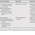
132 Meniscal signal not related to tear is usually secondary to degeneration. In addition, there are numerous interpretive pitfalls related to normal variants, which have been well described.1 A common artifact is the magic angle effect on intermediate and T1-weighted magnetic resonance images (T1WI). This usually involves the posterior horn lateral meniscus, is ill-defined, does not extend to an articular surface, and is not seen on T2-weighted magnetic resonance images (T2WI). Less common causes of increased meniscal signal not related to a tear are Meniscal cysts are associated with a meniscal tear. An intrasubstance meniscal cyst is usually less intense than fluid on T2WI. It resembles intrasubstance degeneration, but the meniscus is swollen.2 On magnetic resonance imaging (MRI), a meniscal ossicle should not be confused with a tear as an ossicle is rounded and has bone signal intensity (high on T1WI and low on T2WI).3
Increased Meniscal Signal Not Related to Tear
Meniscal Cysts
Meniscal Ossicles
Chondrocalcinosis
Related posts:

Stay updated, free articles. Join our Telegram channel

Full access? Get Clinical Tree


