, Lawrence A. Zumo2 and Valerie Sim3
(1)
Parkinson’s Clinic of Eastern Toronto, Toronto, ON, Canada
(2)
Silver Spring, Cheverly, MD, USA
(3)
Centre for Prions and Protein Folding Diseases, University of Alberta, Edmonton, AB, Canada
Abstract
Many types of infectious organisms can affect the nervous system, including bacteria, fungi, viruses and parasites. It is beyond the scope of this reference manual to include every possible infection, but we include a description of imaging findings that are typical for tropical neurological diseases such as cerebral malaria, microsporidiosis, trypanosomiasis, leshmaniasis, dengue fever and snake bite. In addition, we present cases of CMV pachygyria, tuberculosis of the spine (Pott’s disease) and spinal cord (syrinx), and complications of HIV infection such as toxoplasmosis and variable presentations of progressive multifocal leukodystrophy from JC virus.
A review of tropical neurological disease imaging characteristics.
Cerebral malaria: multiple cortical/subcortical contrast enhancing lesions with transtentorial herniation in fatal cases
Microsporidiosis: multiple ring enhancing lesions at the gray/white junction
African trypanosomiasis: basal ganglia hypodensities
CNS leshmaniasis: basal skull and meningeal inflammatory changes, optic nerve involvement, in addition to the more common signs and symptoms of peripheral neuropathy
Dengue fever: intracranial hemorrhage
Snake bite: hemorrhagic and ischemic infarct due to disseminated intravascular coagulation and toxin induced vasculitis
Case 9.1 CMV Pachygyria
A neonate was found to have poor muscle tone (hypotonia), poor muscle control, feeding difficulties, small head circumference and new onset infantile spasms. TORCH panel showed CMV infection. A CT was performed (Fig. 9.1)
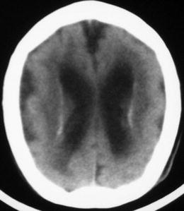

Figure 9.1
Axial CT showing pachygyria and incomplete development of the Sylvian fissures (Image courtesy of M. Koby)
Explanation and Diagnosis
This neonate’s hypotonia, feeding difficulties and poor muscle control as well as new onset infantile spasms can be attributed to intrauterine CMV infection as demonstrated by the TORCH panel. Figure 9.1 shows thickened cerebral cortices with few large gyri with incomplete development of the Sylvian fissures, consistent with pachygyria, a rare developmental disorder resulting from the abnormal migration of neurons in the developing brain and nervous system. This can be seen in other metabolic disorders such as Zellweger syndrome (peroxisome biogenesis disorder).
< div class='tao-gold-member'>
Only gold members can continue reading. Log In or Register to continue
Stay updated, free articles. Join our Telegram channel

Full access? Get Clinical Tree


