Infection imaging has long been an important indication for scintigraphy. Gallium-67 citrate (Ga-67) was the first infection-seeking radiopharmaceutical used clinically. It is still in use today, however, in a much more limited role than in the past. For decades now, In-111 oxine leukocytes have been the primary radiopharmaceutical used to detect infection. Tc-99m hexamethylpropyleneamine oxime (HMPAO)–labeled leukocytes have also found a role. In recent years, Fluorine-18 fluorodeoxyglucose (F-18 FDG) is increasingly being used for confirming or excluding infection ( Box 15.1 ). Single-photon emission computed tomography with computed tomography (SPECT/CT) and positron emission tomography with computed tomography (PET/CT) have added specificity and improved localization for all of the radiopharmaceuticals.
Ga-67 citrate
In-111 oxine–labeled leukocytes
Tc-99m HMPAO–labeled leukocytes
F-18 fluorodeoxyglucose (FDG)
Tc-99m sulesomab (LeuTech; approved in Europe)
HMPAO, Hexamethylpropyleneamine oxime.
Pathophysiology of Inflammation and Infection
Inflammation is a tissue response to injury attracting cells of the immune system to the site of damage. It is triggered by tissue injury (e.g., trauma, foreign particles, or neoplasm). The inflammatory response results in increased blood flow, vasodilation, increased vascular permeability, and migration of leukocytes out of blood vessels into the tissues (chemotaxis). The plasma carries proteins, antibodies, and chemical mediators that modulate the inflammatory response to the site of infection ( Fig. 15.1 ). Leukocytes are attracted to the infection site in response to chemoattractants (e.g., bacterial products). Infection implies the presence of microorganisms. Although infection is usually associated with inflammation, the reverse is not always true. Infection without inflammation occurs in severely immunosuppressed patients.

Leukocytes are the major cellular components of the inflammatory and immune response that protect against infection and neoplasia and assist in the repair of damaged tissue. Nucleated precursor cells differentiate into mature cells within the bone marrow. Peripheral leukocytes include granulocytes (neutrophils 60%, eosinophils 3%, basophils 1%), lymphocytes (30%), and monocytes (5%). Only 2% to 3% of neutrophils reside in the circulating blood, using it transiently when moving to sites of need. The rest are distributed in a “marginated” pool that is adherent to vascular endothelial cells in tissues, mostly in the bone marrow but also in the spleen, liver, lung, gastrointestinal tract, and oropharynx. These marginated cells can be marshaled into the circulating pool by various stimuli, including exercise, epinephrine, or bacterial endotoxins.
Neutrophils respond to an acute inflammatory stimulus by migrating toward an attractant (chemotaxis) and enter tissues between postcapillary endothelial cells (diapedesis). The neutrophils phagocytize the infectious agent or foreign body and enzymatically destroy it within cytoplasmic vacuoles. Lymphocytes, including B cells, T cells, and natural killer cells, arrive at inflammatory sites during the chronic phase. Monocytes act as tissue scavengers, phagocytosing damaged cells and bacteria and detoxifying chemicals and toxins. At sites of inflammation, they transform into tissue macrophages. Corticosteroids and ethanol inhibit the inflammatory process.
Radiopharmaceuticals
In-111-labeled leukocytes were first introduced in 1977. Indium-111 8-hydroxyquinoline (oxine) and Tc-99m hexamethylpropyleneamine oxime (HMPAO)–labeled leukocytes each have advantages and disadvantages ( Table 15.1 ).
| Advantages and Disadvantages | In-111-Oxine WBCs | Tc-99m-HMPAO WBCs |
|---|---|---|
| Radionuclide readily available | No | Yes |
| Stable radiolabel, no elution from cells | Yes | No |
| Allows labeling in plasma | No | Yes |
| Radiation dosimetry | Poor | Good |
| Early routine imaging | No | Yes |
| Half-life allows for delayed imaging | Yes | No |
| Imaging time | Long | Short |
| Permits dual-isotope imaging | Yes | No |
| Intestinal and renal clearance | No | Yes |
| Image resolution | Fair | Good |
Indium-111 Oxine Leukocytes
The radionuclide Indium-111 (In-111) is cyclotron produced, decays by electron capture, emits two gamma photons (173 and 247 keV), and has a physical half-life of 67 hours (2.8 days; Table 15.2 ).
| Radionuclide | Half-Life | Photopeak (keV) | % Photon Abundance |
|---|---|---|---|
| In-111 | 67 hours | 173 | 89 |
| 247 | 94 | ||
| Tc-99m | 6 hours | 140 | 89 |
| F-18 | 110 minutes | 511 | 97 |
| Ga-67 | 78 hours | 93 | 41 |
| 185 | 23 | ||
| 288 | 18 | ||
| 394 | 4 |
Adequate radiolabeling to provide good image quality usually requires 5000/mm 3 peripheral leukocytes, although sometimes the study can be performed with counts as low as 2000/mm 3 . The in vitro labeling procedure requires a minimum of 2 hours. Lacking facilities and trained personnel to radiolabel the cells, imaging clinics send a patient’s blood to an outside commercial radiopharmacy for radiolabeling. The time required for sending the blood to the radiopharmacy, radiolabeling the cells, returning the cells to the clinic, and reinfusing them is 3 to 4 hours.
Fifty milliliters of venous blood usually ensures sufficient leukocytes for adequate labeling; 20 to 30 mL may be acceptable for children. Radiolabeling must be performed under a laminar flow hood to ensure sterility. Careful handling is required to avoid damaging cells, which could adversely affect their migration and viability. Proper labeling does not affect normal physiological function. The radiolabel remains stable in vivo for more than 24 hours.
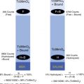
The methodology for radiolabeling leukocytes is described in detail in Box 15.2 . Radiolabeling cannot be performed in plasma, so it must be separated and retained for later resuspension with the leukocytes before reinfusion. The leukocyte pellet is suspended in saline and incubated with In-111 oxine. The lipid solubility of the In-111 oxine complex allows it to diffuse through cell membranes. Intracellularly, the complex dissociates, oxine diffuses back out of the cell, and In-111 binds to nuclear and cytoplasmic proteins. In-111 oxine binds to granulocytes, lymphocytes, monocytes, platelets, and erythrocytes. Pure In-111 granulocyte preparations have been radiolabeled and used clinically. However, they require density gradient centrifuge separation techniques and have not shown a clinical advantage over mixed leukocytes. They are generally not used clinically.
Preparation
- 1.
Patient’s peripheral leukocyte count should be >5000 cells/mm 3 .
Procedure
- 1.
Collect autologous blood.
Draw 30 to 50 mL into an anticoagulant citrate dextrose (ACD) anticoagulated syringe using a 19-gauge needle.
- 2.
Isolate leukocytes:
Separate red blood cells (RBCs) by gravity sedimentation and 6% Hetastarch, a settling agent.
Centrifuge the leukocyte-rich plasma at 300 to 350 g for 5 minutes to remove platelets and proteins.
A leukocyte button forms at the bottom of the tube.
Draw off and save the leukocyte-poor plasma (LPP) for later washing and resuspension.
- 3.
Label leukocytes:
Suspend leukocytes (LPP) in saline.
Incubate with In-111 oxine for 30 minutes at room temperature and gently agitate.
Remove unbound In-111 by centrifugation. Save wash—calculate labeling efficiency.
- 4.
Prepare injectate:
Resuspend 500 μmCi In-111-labeled leukocytes in saved plasma (LPP).
Inject via peripheral vein within 2 to 4 hours.
- 5.
Perform quality control:
Microscopical examination of cells
Calculate labeling efficiency:
Assay the cells and wash in dose calibrator.
Labeling efficiency = C/([C + W] × 100%)
where C is activity associated with the cells, and W is activity associated with the wash.
Standard quality-control measures (e.g., testing for sterility and pyrogenicity) cannot be performed because of the need for prompt reinfusion after labeling to ensure cell viability. However, the final radiopharmaceutical preparation should be examined for abnormal morphology, clumping, excessive red blood cell (RBC) contamination, and percent labeling efficiency, typically ranging from 75% to 90%. When labeling is less than 50%, the cells should not be administered to the patient. The final preparation contains radiolabeled granulocytes, lymphocytes, and monocytes and some platelets and erythrocytes.
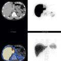
After infusion of the In-111 leukocytes, no significant elution of the In-111 from the leukocytes occurs. Initial distribution is to the blood pool, lungs, liver, and spleen. Localization is the result of diapedesis and chemotaxis ( Table 15.3 ). Lung uptake is caused by activation from in vitro cell manipulation, which may be increased by the underlying disease process. By 4 hours after reinjection, lung and blood-pool activity markedly decrease ( Fig. 15.2 ). By 24 hours, the greatest uptake is in the spleen, followed by the liver and bone marrow. Persistent blood pool suggests a high percentage of labeled RBCs. Genitourinary, hepatobiliary, and intestinal clearance are not normally seen. The ultimate test of the viability of leukocytes is in vivo function manifested by a normal distribution within the body and the ability to detect infection ( Table 15.4 ). Uptake outside of expected normal uptake sites suggests infection ( Figs. 15.3 and 15.4 ). If the infused white blood cells (WBCs) become nonviable, increased liver and lung uptake is seen.
| Radiopharmaceutical | Mechanism |
|---|---|
| Radiolabeled leukocytes | Diapedesis and chemotaxis |
| F-18 fluorodeoxyglucose | Glucose metabolism |
| Ga-67 citrate | Iron binding to lactoferrin and bacterial siderophores |

| Radiopharmaceutical | Liver | Spleen | Marrow | Bone | Gastrointestinal | Genitourinary | Lung |
|---|---|---|---|---|---|---|---|
| Ga-67 citrate | ∗∗∗ | ∗ | ∗ | ∗ | ∗∗∗ | ∗ | |
| In-111 oxine–labeled leukocytes | ∗∗ | ∗∗∗ | ∗∗ | ∗ | |||
| Tc-99m HMPAO–labeled leukocytes | ∗∗ | ∗∗∗ | ∗∗ | ∗∗ | ∗∗ | ∗ | |
| F-18 FDG | ∗∗∗ | ∗∗ | ∗ | ∗∗ | ∗∗ |


An imaging protocol for In-111 oxine leukocytes is described in Box 15.3 . Images are acquired on their 173- and 247-keV photopeaks (20% window) using a medium-energy collimator. Whole-body imaging is routine. High-count spot images, SPECT, and SPECT/CT are options. The spleen receives the highest radiation dose, precluding its use in pediatric patients. Dosimetry is detailed (see the Appendix). Images are routinely acquired 24 hours after reinfusion ( Table 15.5 ). Further delayed images rarely give additional information. Early imaging (e.g., at 4 hours) is less sensitive than 24-hour imaging for detecting infection, although when the patient is quite sick, early imaging may detect the focus of infection (e.g., an abscess that requires urgent intervention). Four-hour imaging is standard for inflammatory bowel disease. Inflamed mucosal cells slough, become intraluminal, and move distally. Thus, 24-hour images can be misleading and erroneous as to the site of inflammation. Scintigraphic images reflect the distribution of leukocytes in the body. Focal or diffuse activity outside their expected normal distribution suggests inflammation or infection (see Table 15.4 ).
Radiopharmaceutical
In-111 oxine in vitro–labeled leukocytes, 500 μCi (18.5 MBq)
Instrumentation
Camera: Large field of view
Window: 20% centered over 173- and 247-keV photopeaks
Collimator: Medium energy
Patient Preparation
Draw 50 mL of blood. Radiolabel white cells (see Box 15.2 ).
Procedure
Inject labeled cells intravenously, preferably by direct venipuncture through a 19-gauge needle. Contact with dextrose in water solutions may cause cell damage.
Imaging at 4 hours is critical in localizing inflammatory bowel disease.
Perform routine whole-body imaging at 24 hours.
Acquire spot views as needed: anterior abdomen for 500k counts, then other images for equal time. Include anterior and posterior views of the chest, abdomen, and pelvis and spot images of specific areas of interest (e.g., feet) for a minimum of 200k counts or 20 minutes.
Perform SPECT or SPECT/CT as needed.
SPECT, Single-photon emission computed tomography; SPECT/CT, single-photon emission computed tomography with computed tomography.
| Radiopharmaceutical | Time (hours) |
|---|---|
| Ga-67 citrate | 48 |
| In-111-oxine leukocytes | 24 |
| Tc-99m-HMPAO leukocytes | 1-4 |
| F-18 fluorodeoxyglucose | 1 |
Interpretive pitfalls (i.e., potential false-positive and false- negative findings) should be kept in mind ( Box 15.4 ). Leukocytes accumulate at sites of inflammation (e.g., intravenous catheters; nasogastric, endogastric, and drainage tubes; tracheostomies; colostomies; and ileostomies). Unless intense, this uptake should be considered normal. Uninfected postsurgical wounds show uptake for 2 to 3 weeks. If uptake is intense, persists, or extends beyond the surgical wound site, infection should be suspected ( Fig. 15.5 ). Uptake occurs at healing bone-fracture sites, with the amount depending on the length of time since fracture, its severity, and whether it is healing properly.
False-Negative Results
Vertebral osteomyelitis
Chronic low-grade infection
Parasitical, mycobacterial, or fungal infections
Hyperglycemia
Corticosteroid therapy
False-Positive Results
Gastrointestinal bleeding
Healing fracture
Swallowed leukocytes; oropharyngeal, esophageal, or lung disease
Surgical wounds, stomas, or catheter sites
Hematomas
Tumors
Accessory spleens
Renal transplant
Pseudoaneurysm

A major advantage of In-111 leukocytes is the normal lack of intraabdominal activity. However, on occasion, abdominal activity may not be due to infection but rather the result of an accessory spleen, splenosis, pseudoaneurysm, or a noninfected hematoma ( Figs. 15.6 and 15.7 ). Renal transplants normally accumulate leukocytes, probably as a result of low-grade rejection ( Fig. 15.8 ). Intraluminal intestinal activity can result from swallowed or shedding cells that occur with pharyngitis, sinusitis, esophagitis, pneumonia, or gastrointestinal bleeding.


The sensitivity of leukocyte imaging may be reduced in conditions that alter leukocyte function (e.g., hyperglycemia, steroid therapy, and chemotherapy). Conflicting data exist regarding the sensitivity of In-111 leukocyte scintigraphy for detecting infection in patients receiving antibiotics. Investigations have not found a difference in sensitivity for detection of acute versus chronic infections. Although chronic inflammations preferentially attract lymphocytes, monocytes, plasma cells, and macrophages, they still have significant neutrophilic infiltration.
Tc-99m-HMPAO Leukocytes
The radionuclide technetium-99m (Tc-99m) is generator-produced from molybdenum-99. Tc-99m decays by isomeric transition and emits one gamma photon of 140 keV, with a 6-hour physical half-life (see Table 15.2 ). The radiopharmaceutical Tc-99m exametazime (Ceretec) is labeled to leukocytes by HMPAO. Tc-99m HMPAO was originally approved by the U.S. Food and Drug Administration (FDA) for cerebral perfusion imaging. Its lipophilicity allows it to cross cell membranes. In brain imaging, it crosses the blood–brain barrier and is taken up by cerebral cortical cells, where it changes into a hydrophilic complex and becomes trapped. This led to the use of Tc-99m HMPAO to label leukocytes. The FDA has accepted Tc-99m HMPAO–labeled leukocytes as an acceptable alternative use of Tc-99m HMPAO.
Tc-99m HMPAO–labeled leukocytes have some advantages over In-111-labeled leukocytes (see Table 15.1 ). The radiation dose to the patient is considerably lower (see the Appendix); thus, greater activity can be administered, resulting in higher photon yield and better image quality.
The methodology for radiolabeling leukocytes with Tc-99m HMPAO is similar to that for In-111 oxine labeling of leukocytes (see Box 15.2 ). The differences are that Tc-99m-HMPAO leukocyte labeling can be performed in plasma and that the resulting labeling is primarily of granulocytes. The biological half-life of Tc-99m-HMPAO leukocytes in blood is shorter than that of In-111 oxine leukocytes, 4 versus 6 hours, because of slow elution of the Tc-99m HMPAO from circulating radiolabeled cells. Tc-99m-HMPAO leukocytes distribute in the body similar to In-111-labeled cells, except that the Tc-99m leukocytes have hepatobiliary and genitourinary clearance due to elution from the leukocytes ( Fig. 15.9 ; see also Table 15.4 ). In addition, a secondary labeled hydrophilic complex is excreted, also seen with Tc-99m-HMPAO brain imaging. The kidneys and bladder are normally visualized by 1 to 2 hours after injection. Biliary and bowel clearance occurs as early as 2 hours, is routinely seen by 3 to 4 hours, and increases with time ( Fig. 15.10 ; see also Fig. 15.9 ).


An important advantage of Tc-99m-HMPAO leukocytes is its much lower radiation dose to the spleen, particularly in children, <2.0 rads compared with approximately 50 rads for In-111 leukocytes (see the Appendix). Thus, Tc-99m-labeled leukocytes are not indicated for children. Because of the shorter physical half-life and hepatobiliary and urinary clearance of Tc-99m-HMPAO leukocytes, images are acquired at an earlier time point than In-111 oxine–labeled WBCs (see Table 15.5 ). An imaging protocol is described in Box 15.5 . Abdominal imaging should be performed approximately 2 hours after reinfusion to minimize the likelihood of urinary and gastrointestinal clearance. For other body regions, such as the chest and extremities, later imaging at 3 to 4 hours is recommended, although further delayed imaging may at times be useful ( Fig. 15.11 ). Blood-pool activity is commonly seen with Tc-99m leukocytes compared with In-111-labeled leukocytes because of the elution of activity from the radiolabeled cells. This can potentially complicate interpretation in the chest or major vessels. Care should be used in the interpretation of intraabdominal activity because of the normal genitourinary and hepatobiliary clearance. Potential false-positive and false-negative results are listed in Box 15.4 . Delayed imaging, particularly SPECT/CT, can often improve localization.
Patient Preparation
Wound dressings changed before imaging
Radiopharmaceutical
Tc-99m-HMPAO in vitro–labeled WBCs, 10 mCi (370 MBq)
Instrumentation
Camera: Large field of view; two-headed camera preferable for whole-body imaging
Collimator: Low energy, high resolution
Windows: 20%, centered over 140-keV photopeaks
Patient Preparation
Draw 50 mL of blood.
Procedure
Radiolabel patient’s leukocytes in vitro with Tc-99m HMPAO.
Reinject labeled cells intravenously, preferably by direct venipuncture through 19-gauge needle. Contact with dextrose in water solutions may cause cell damage.
Imaging
Imaging by 2 hours is necessary for intraabdominal imaging or to localize inflammatory bowel disease.
Imaging at 4 hours or later may be advantageous for peripheral skeleton (e.g., osteomyelitis of feet).
Whole-body imaging: Two-headed camera with whole-body acquisition for 30 minutes; 10-minute spot images for regions of special interest
SPECT or SPECT/CT in selected cases.
HMPAO, Hexamethylpropyleneamine oxime; SPECT, single-photon emission computed tomography; SPECT/CT, single-photon emission computed tomography with computed tomography; WBC, white blood cell.
Related posts:

Stay updated, free articles. Join our Telegram channel

Full access? Get Clinical Tree


