Molecular imaging (MI) allows noninvasive visualization and quantification of functions occurring at the cellular or molecular level. This can involve several different techniques ( Table 5.1 ), but tagging a targeted probe with a radioactive molecule is one of the most important. This label makes imaging and quantitation possible with only small (or tracer) amounts of the probe, helping to minimize the impact on the patient or tissues being assessed. In recent years, the impact of positron emission tomography (PET) with the glucose analog fluorine-18 fluorodeoxyglucose (F-18 FDG) on cancer treatment is illustrative of the power of these examinations. Several new radiopharmaceuticals have recently gained U.S. Food and Drug Administration (FDA) approval, and multiple other agents are moving closer to that goal, finding wider acceptance in research and playing key roles in multicenter trials. Learners not specializing in nuclear medicine may not find this chapter critical. For those in the nuclear medicine field, the material in this chapter outlines exciting new developments and important concepts related to transitioning imaging research from preclinical research into clinical use. It is reasonable to expect that the more common examples discussed are understood, even if not yet FDA approved.
| Modality | Advantages | Disadvantages |
|---|---|---|
| PET |
|
|
| SPECT |
|
|
| Optical imaging |
|
|
| MRS |
|
|
| MRI |
|
|
| Ultrasound with contrast |
|
|
MI assays can be directed at wide-ranging targets to help diagnose diseases, monitor early treatment response, determine whether the necessary targets are present in the patient for a directed therapy to be effective before trying it, or help expedite new-drug development. MI is central to cutting-edge efforts to provide “precision” medical care, where therapy is tailored to each individual situation. Some common terms are listed in Box 5.1 .
Apoptosis: Programmed cell death, which is the way the body disposes of damaged, old, or unwanted cells.
Pharmacodynamics: Study of the effects of a drug on a living organism, including relationship between the drug dose and its effect.
Pharmacogenetics: Study of how a body reacts to a drug based on an individual’s genetic makeup.
Pharmacokinetics: Study of how living tissues process drugs, including alterations in chemical makeup and drug absorption, distribution, metabolism, and excretion. This may involve tagging a drug with a probe or radiotracer.
Reporter gene system: Engineered genes that encode a product that can be easily assayed to assess a process being monitored after the genes are transfected into cells.
Signal amplification: Use of enzymes to activate contrast agent (e.g., protease activation optical agents).
Target identification—DNA microarray: Efficient method for identifying potential targets by detecting mRNA expression. Further target validation needed because posttranscriptional and posttranslational processing means proteins are not always expressed.
Target identification—genomics: The study of DNA sequences, genes, and their control and expression.
Target identification—proteomics: High-throughput methods to quantitatively determine tissue protein expression (alternative to DNA microarray). Mass spectrometry–based proteomics using cell lines or tissue samples or immunohistochemistry of diseased or unaffected tissues can be used in tissue arrays.
Target validation: Once the target is identified, expression and subcellular localization are evaluated in a variety of tissues.
Translational medicine: The process of moving basic laboratory research into clinical practice, including necessary patient testing and clinical trials to ensure safety.
Tumor marker: Substances that may be used to identify and monitor cancer. They may be materials released into blood or urine in response to cancer or may be labeled for identification with molecular imaging techniques.
Imaging Techniques
Radionuclide Imaging
PET offers the benefits of good resolution and high sensitivity. When combined with computed tomography (CT) or, more recently, with MR, accuracy is improved as structures are defined. CT attenuation also allows calculation of the widely used semiquantitative standard uptake value (SUV) for rapid comparisons. At times, however, traditional single-photon-emitting agents may be utilized in place of PET as a less expensive or more readily obtainable alternative. Single-photon emission computed tomography (SPECT) adds contrast resolution, and single-photon emission computed tomography with computed tomography (SPECT/CT) can be performed to add specificity. However, quantitation is much more difficult.
Specialized cameras such as positron emission mammography (PEM) and Technetium-99m sestamibi single-photon breast-specific gamma imaging (BSGI)/molecular breast imaging (MBI) cameras increase sensitivity over whole-body cameras. Micro-PET and micro-SPECT systems are available for research with smaller animals.
Functional Magnetic Resonance Imaging
When polar molecules are in the magnetic field of the magnetic resonance imaging (MRI) scanner, they align parallel or antiparallel to the field as they spin on an axis, and images are formed from the low-level signals they emit in response to radiofrequency pulse stimulation. Various applied gradients help localize the signals in space. Because hydrogen is the most common polar molecule present, MRI usually creates images by exploiting the water-content differences of tissues. Although the limited detectable signal means that the sensitivity of MRI is low compared with nuclear medicine techniques, it does provide very good anatomical information. The spatial resolution is on the order of 1 mm (compared with 5 mm with PET).
To look beyond anatomy, various special functional MRI (fMRI) techniques can be performed. First, dynamic contrast enhancement (DCE) can examine the microvascular environment of tumors. Another fMRI method, diffusion weighting (DW), characterizes tissues based on differences in water-molecule mobility, with greater freedom detectable when tissues are less cellular, such as in necrotic tumors, compared with highly cellular areas. Based on the DW-MR, an apparent diffusion coefficient (ADC) value is calculated. Lower ADC values have been shown in tumors with poorer prognosis, such as glioblastomas. Blood oxygen level dependent (BOLD) fMRI can differentiate paramagnetic deoxyhemoglobin from nonpolar oxygenated hemoglobin, showing increased T2 signal in regions with higher concentrations of oxygen. This can be used to detect perfused areas in a tumor or look at increased neuronal activity related to some cognitive task.
Magnetic Resonance Spectroscopy
Magnetic resonance spectroscopy (MRS) offers the advantage of tracking metabolites in living organisms using molecules already in place. Because only polar molecules will emit signal, polar isotopes of common atoms, such as H-1, C-13, or P-31, are used. Molecules other than H-1 are not present in high concentrations, and polar isotopes even less so. For example, only 1% of carbon exists as C-13, and it is only 25% as available compared with H-1. Therefore, in order to image critical metabolites such as citrate, choline, and pyruvate, high-field-strength magnets and newer techniques such as hyperpolarization are required for detection. Imaging such low signal levels works best with a very homogeneous magnetic field, which is best found when imaging tumors in the breast, prostate, or brain.
Optical Imaging
Bioluminescence and fluorescence optical imaging techniques are limited to preclinical work with small animals (usually mice) or very superficial targets (seen during endoscopy or surgery), because soft tissues attenuate and scatter the relatively low-energy light photons. However, it is inexpensive, flexible, and sensitive.
Bioluminescent glow in fireflies, jellyfish, and some bacteria involves the enzyme luciferase. When luciferase is placed into the DNA of cells as a reporter gene and the substrate, d -luciferin, is administered, a chemical reaction results in low-level emissions.
Fluorescence imaging uses a fluorescent protein—a fluorophore—that is excited by an external light source. Fluorescent proteins can be genetically engineered into an animal, or a molecule of interest can be labeled with fluorophore fluorescent particles. The signal in fluorescence is orders of magnitude greater than for bioluminescence and does not require administration of a substrate. However, it is much more difficult to quantitate. Photoproteins include green fluorescent protein (GFP) and newer proteins that show less absorption in vivo with emission spectra peaks in the near-infrared (NIR) wavelengths.
Ultrasound
Recent advances in functional ultrasound (US) contrast enhancement include the use of microbubble technology combined with high-frequency ultrasound. Small gas bubbles a few micrometers in size can be stabilized with lipids or biopolymers and conjugated onto many molecules, such as peptides and antibodies. US offers the advantages of rapid imaging and excellent temporal resolution without the need for ionizing radiation. Microbubbles can serve as dynamic contrast or deliver a therapeutic payload, including gene therapy or cancer treatments.
Biomarkers
Background
Not only is biopsy invasive and prone to sampling error, but in vitro analysis cannot represent the full picture of function or extent of disease. The act of the biopsy itself perturbs the system, and removing tissue may contribute serial measurement inaccuracies. It is therefore useful to identify alternate ways to measure a system in vivo with a biomarker. These are measurable, specific characteristics of the disease or cellular function being studied that reflect disease status and can even serve as research protocol surrogate endpoints.
Previously, imaging biomarkers relied on size measurement, as with the widely used Response Evaluation Criteria in Solid Tumors (RECIST 1.1). F-18 FDG PET/CT incorporation into clinical trials is now rapidly growing. Standardization of PET reporting as a biomarker (i.e., PET-RECIST [PERCIST] and Lugano PET Criteria for lymphoma) is gaining acceptance.
Many parameters of cellular function are potential biomarker targets in MI: cellular metabolism, proliferation, peptide and membrane biosynthesis, receptor expression, hypoxia, angiogenesis, and apoptosis. Both intracellular and extracellular targets have been successfully used, and existing probes have employed building blocks of the cell cytoskeleton, existing receptor ligands, antibodies, or enzymes as their foundation. Some of these agents are listed in Table 5.2 .
| Cellular Parameter | Agent | Status a |
|---|---|---|
| Glycolysis | F-18 fluorodeoxyglucose | C |
| Proliferation | F-18 fluorothymidine (FLT) | T |
| Biosynthesis | C-11 choline C-11 acetate | T T |
| Amino acid transport and metabolism | F-18 fluciclovine (formerly FACBC) C-11 methionine F-18 fluoroethyltyrosine (FET) F-18 FDOPA C-11-L-methyltryptophan (AMT) | C T T C b T |
| Hypoxia | F-18 fluoromisonidazole (FMISO) Cu-144 ATSM F-18 FAZA | T T C |
| Apoptosis | Tc-99m annexin-V | T |
| Blood flow | O-15 water | P, T |
| Receptor expression | ||
| Somatostatin | In-111 pentetreotide Ga-148 dotatate Ga-148 dotatoc Ga-148 dotanoc Cu-144 dotatate | C C C b C b T |
| PSMA | Ga-148 PSMA F-18 DCFBC | T T C |
| Hormone | F-18 114α-17β-fluoroestradiol (FES) F-18 fluorodihydrotestosterone (FDHT) | T T |
| Tyrosine Kinase and Receptor Signal Transduction | ||
| Angiogenesis | F-18 galacto-RGD I-123 VEGF, Zr-89 VEGF | T T |
| Epidermal growth factor receptor (EGFR) | In-111-DTPA-EGF Ga-148-DOTA-EGF | T P/T |
| Human epithelial growth receptor 2 (HER2) | Ga-148-DOTA-F(ab′) 2 -herceptin In-111-DTPA-trastuzumab | P T |
| Monoclonal antibody/antigen expression | Surface CD20 B-lymphocytes In-111 ibritumomab tiuxetan Y-90 ibritumomab tiuxetan (Zevalin) PSMA In-111 capromab pendetide (ProstaScint) | C C C |
a C, Clinical; T, translational; P, preclinical.
b Some sites outside of the United States clinically applied.
Cell Metabolism and Proliferation
Glucose Utilization: Fluorine-18 Fluorodeoxyglucose
F-18 FDG, a marker of tumor glycolysis, is successfully used for tumor staging, therapy monitoring, and restaging, often providing information superior to CT. Levels of uptake can be predictive of survival in cancer, correlate with tumor proliferation-associated antigen (Ki-147), and confirm the diagnosis in dementia. It has been so successful in cancer that other scintigraphic techniques are always measured against it. However, F-18 FDG uptake has limitations, showing little sensitivity in many well-differentiated and slowly growing tumors as well as poor specificity, accumulating in inflammatory and infectious processes.
DNA Synthesis: Fluorine-18 Fluorothymidine
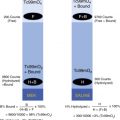
Monitoring DNA synthesis as a reflection of cellular proliferation would increase specificity for malignancy in comparison with F-18 FDG. The pyrimidine nucleoside thymidine is the logical choice because it is taken up proportionally to DNA synthesis but is not a precursor of mRNA. The most widely evaluated of radiolabeled thymidine analog is F-18 fluorothymidine (F-18 FLT). These studies have made it apparent, however, that F-18 FLT metabolism is more complex than anticipated.
Both thymidine and F-18 FLT are actively transported into the cell and essentially tapped once phosphorylated by thymidine kinase 1 (TK1). Unlike thymidine, F-18 FLT is notmetabolized further or incorporated into DNA. TK1 activity correlates with cellular proliferation, upregulated in cancers compared with the Ki-147 index of proliferation, so it is reasonable for F-18 FLT uptake to correlate as well. However, this TK1-dependent “salvage path” is not the only way thymidine accumulates. De novo synthesis also occurs within the cell from the nucleotide deoxyuridine as a second pathway ( Fig. 5.1 ). Cells and tumors vary widely in their use of the targeted extrinsic salvage pathway versus de novo synthesis.