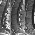
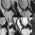
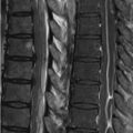
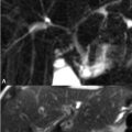
86 Insufficiency Fractures, Acetabular Labrum Fractures characteristically demonstrate hyperintensity on STIR and FS T2WI as well as linear hypointensity on T1WI perpendicular to the direction of force. A left pubic insufficiency fracture is illustrated on the T1WI and FS T2WI of Figs. 86.1A,B, respectively. The left parasymphysial area of the pubis exhibits low SI on (A) T1WI with a more lateral linear band of hypointensity, perpendicular to the direction of weight-bearing. (B) FS T2WI demonstrates hyperintensity within this region, further suggesting an insufficiency fracture. Bone contusion may appear similar, but differs in clinical history and also in the lack of linear-appearing hypointensity. Figure 86.2A,B illustrates a sacral insufficiency fracture on the coronal T1WI and axial FS T2WI, respectively. Vertically oriented linear hypointensity is present within the right sacrum on the former, consistent with an insufficiency fracture. Sacral fractures are often present on lumbar spine MRI, although they may be missed due to location at the periphery of the region of interest. Careful examination of sagittal images for such findings is therefore warranted.
Stay updated, free articles. Join our Telegram channel

Full access? Get Clinical Tree


