Interspinous Bursitis (Baastrup’s Disease)


Clinical Presentation


The patient is a 75-year-old male who presented with severe mid low back pain. He has difficulty walking distances but reports no lower extremity pain. Physical examination reveals some vague tenderness to deep palpation of the back. No lower extremity weakness. Normal sensory examination. Normal deep tendon reflexes.


Imaging Presentation


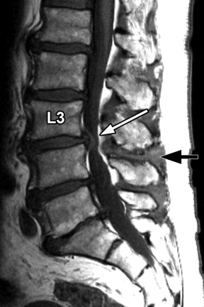
Multilevel spondylosis deformans, intervertebral disc degeneration, and generalized disc bulging are seen with imaging. Severe circumferential thecal sac narrowing is present at the L3-4 level as a result of disc bulging, thickened ligamentum flava, and bilateral facet hypertrophy. Abnormal T2 signal hyperintensity and contrast enhancement is demonstrated in the L3-4 interspinous ligaments and adjacent spinous processes, and contrast enhancement of the L3-4 facet capsules is shown bilaterally. Findings are consistent with severe central stenosis at the L3-4 level, active inflammatory facet arthropathy, and interspinous ligamentous inflammation (i.e., interspinous bursitis, also known as Baastrup’s disease ) ( Figs. 39-1 to 39-7 ) .


Figure 39-1


Spinal Stenosis, Interspinous Bursitis (Baastrup’s Disease).

Sagittal T1-weighted MRI reveals severe central canal stenosis at L3-4 level ( white arrow ) due to disc bulging and ligamentum flavum thickening. The L3-4 interspinous ligament is thickened ( black arrow ).


Figure 39-2


Spinal Stenosis, Interspinous Bursitis.

Corresponding sagittal fat-saturated sagittal MRI in same patient as in Figure 39-1 . T2 hyperintensity in L3-4 interspinous tissues and adjacent L3 and L4 spinous processes ( large white arrows ). Central canal stenosis indicated by small white arrow .


Figure 39-3


Spinal Stenosis, Interspinous Bursitis.

Corresponding sagittal contrast-enhanced T1-weighted fat-saturated sagittal image in same patient as in Figures 39-1 and 39-2 . There is narrowing of the interspinous space and enhancing tissue in the interspinous space associated with small erosions of the adjacent spinous processes ( arrows ).


Figure 39-4


Spinal Stenosis, Interspinous Bursitis.

Same patient as in Figures 39-1 to 39-3 . Contrast-enhanced parasagittal T1-weighted fat-saturated sagittal MR image reveals enhancing tissue ( arrows ) in the interspinous and paraspinous regions consistent with inflammation of the interspinous ligaments and interspinous bursae.


Figure 39-5


Spinal Stenosis, Interspinous Bursitis.

Corresponding axial T1-weighted fat-saturated sagittal MRI in same patient as in Figures 39-1 to 39-4 , at level of L3 spinous process. Enhancing tissue ( arrows ) is present on either side of the spinous process ( S ) is consistent with enhancement of paraspinous ligaments and/or margins of interspinous bursae. There are 2 ovoid erosions within the spinous process ( S ) containing enhancing tissue.


Figure 39-6


Spinal Stenosis, Interspinous Bursitis.

Corresponding axial T1-weighted fat-saturated sagittal MR image in same patient as in Figures 39-1 to 39-5 , at L3-4 intervertebral disc level. Heterogeneous enhancement of the interspinous ligaments and paraspinous tissues is demonstrated ( arrows ).


Figure 39-7


Spinal Stenosis, Interspinous Bursitis.

Corresponding axial T1-weighted fat-saturated sagittal image in same patient as in Figures 39-1 to 39-6 , at level of L4 spinous process. Enhancement of parafacetal soft tissues ( arrows ) is consistent with active inflammatory facet arthropathy (facet synovitis).


Discussion


The space between adjacent lumbar spinous processes contains an interspinous ligament or ligaments and bilateral paraligamentous bursae. Small, short bilateral paired muscles, the interspinous lumborum muscles, are located on both sides of the interspinous ligament at a given lumbar level. These tissues can become inflamed and be a source of back pain. Interspinous ligamentous inflammation or bursitis (often referred to as Baastrup’s disease or Baastrup’s sign ) most commonly occurs at the L4-5 level but can occur at any lumbar level.


Normally, the spinous processes are covered by noncartilaginous connective tissue. As this disorder evolves, chronic active or episodic inflammation occurs in the interspinous ligaments, and over time the abutting surfaces of the involved spinous processes contact and eventually become flattened and sclerotic, with small cystic erosions or geodes occurring where the spinous processes contact ( Figs. 39-8 to 39-10 ) . This process may lead to interspinous adventitial bursal formation and eventually formation of a synovial-lined articulation between the spinous processes. When interspinous bursal formation occurs, both calcium pyrophosphate dihydrate (CPPD) and hydroxyapatite crystal deposition may be present in the bursa. When interspinous ligamentous inflammation occurs, the adjacent posterior paraspinal musculature and nearby facet capsules may also be inflamed ( Figs. 39-11 to 39-13 ) . Patients with interspinous ligamentous inflammation are usually symptomatic, reporting moderate to intense low back pain near the midline.


Figure 39-8


Interspinous Bursitis.

67-year-old female with chronic low back pain, intervertebral disc degeneration, and Baastrup’s disease. Sagittal CT image showing large L5-S1 bulging disc ( BD ) and intervertebral disc narrowing associated with vacuum disc phenomenon ( small arrows ). The L5-S1 interspinous space markedly narrowed and there are bone erosions and sclerosis where the spinous processes contact each other ( arrows ).


Figure 39-9


Interspinous Bursitis.

Corresponding sagittal T2-weighted MRI in same patient as in Figure 39-8 . T2 hyperintense tissue is demonstrated in the L4-5 interspinous tissues ( arrow ). Note multilevel central canal stenosis.

Aug 25, 2019 | Posted by in NEUROLOGICAL IMAGING | Comments Off on Interspinous Bursitis (Baastrup’s Disease)

Full access? Get Clinical Tree

Get Clinical Tree app for offline access