Interventional and therapeutic procedures

Chapter 34 Interventional and therapeutic procedures


Vascular interventional procedures


Indications


The most common indication for interventional vascular procedures is limb ischaemia, usually of the lower limb. This can present in a variety of ways, such as intermittent claudication (pain in the limb on exercise and relieved by rest), chronic critical limb ischaemia (e.g. causing rest pain, ulcers or gangrene) or acute limb ischaemia (causing pallor, coldness and numbness of the limb).


The common feature is the presence of stenoses or occlusions in the arteries supplying blood to the limb. In general, the greater the severity of the vascular disease, the greater the severity of the symptoms. Limb ischaemia considered suitable for management by interventional radiological techniques can be treated in a variety of ways, such as angioplasty, stent insertion or thrombolysis. It is important to remember, however, that treatment is chosen on the basis of the symptoms, not simply the angiographic appearance.


Embolisation, on the other hand, may be undertaken for a variety of reasons. First, there may be uncontrolled bleeding: for example from the gastrointestinal tract, tumours in various sites and from the kidney, liver or other solid organs after trauma or biopsy. Embolisation is also useful in the treatment of some aneurysms. This is particularly true of aneurysms in the cerebral circulation, where coil embolisation may be used as an alternative to surgical aneurysm clipping to prevent recurrence of subarachnoid haemorrhage. Other indications include treatment of arteriovenous malformations and embolisation of the testicular vein for varicocoele. Uterine artery embolisation can be used for the treatment of uterine fibroids.


Stent grafting is used in the treatment of aneurysms. A true aneurysm describes a situation where the vessel is abnormally dilated because of expansion of all three layers of the vessel wall, making it prone to rupture. When such an aneurysm is present in the abdominal aorta, rupture causes bleeding and without emergency surgery is fatal. Therefore, if an abdominal aortic aneurysm measuring 5.5 cm or more in diameter is identified, it is usual for surgical aneurysm repair to be undertaken to remove the risk of rupture if the patient is sufficiently fit to undergo such major surgery. Since the 1990s, stent grafts have been, and continue to be, developed; this can provide an alternative to open surgical repair, particularly in patients who are at high risk from open surgery. True aneurysms may also arise at other sites, such as the thoracic aorta and iliac arteries, and may also be amenable to treatment by stent grafting.


A false aneurysm is not surrounded by normal vessel wall. Instead, it represents the persistent leakage of blood into a cavity surrounded by haematoma. These are most commonly seen as a result of trauma to the vessel, often iatrogenic, but may also arise due to erosion by tumours or the presence of infection in the vessel wall. Stent grafting may also be useful in the treatment of false aneurysms. However, if infection is thought likely to be present then a stent graft should be avoided if possible, as being of a foreign material its presence would make the infection impossible to eradicate. In deciding whether or not to use a stent graft the site of the false aneurysm should also be considered. For example, a false aneurysm arising from the common femoral artery (CFA) after arterial puncture is positioned directly over the hip joint: a stent graft implanted at this site would be subject to repeated stress and would eventually fail. False aneurysms at this site are therefore better treated with US-guided injection of thrombin, which thromboses the false aneurysm.


Angioplasty


The basic principles of angioplasty are the same in whichever vascular territory they are to be applied. These will be described first, followed by important caveats with respect to different arterial territories.


Once an arterial stenosis requiring treatment has been identified, it is traversed with a suitable guide wire and catheter combination. In very narrow stenoses, which can be very difficult to cross, it can be extremely helpful to use the ‘roadmap’ facility available on modern digital subtraction angiography (DSA) equipment. This allows contrast to be injected while screening, and the image of the vessels to be retained on the monitor. When the screening pedal is next depressed the image of the vessel remains superimposed over the real-time image of the catheter and guide wire as they are being manipulated.


Once the lesion has been crossed it is important that either a guide wire or a catheter should remain across it at all times until the procedure has been completed. When an angioplasty is undertaken, complications such as vessel dissection, occlusion due to acute thrombosis or distal embolisation, or even vessel rupture may occur. If a guide wire has been left across the lesion it is a comparatively simple matter to go on to manage the complication appropriately. If the guide wire has been removed it may be possible to cross the lesion again, but this is often highly complex, is not always successful, and may result in vessel dissection and irretrievable occlusion. At the very least time will be taken up in crossing the lesion again, which in an acute situation is highly counterproductive.


Angioplasty itself is undertaken using a balloon catheter designed for the purpose. Balloons are available in a wide variety of diameters and lengths to suit the vessel and lesion being treated. The majority of balloons have radio-opaque markers at each end to facilitate the correct positioning of the device in relation to the stenosis (some have a marker in the middle). The balloon catheter is inserted through the vascular sheath over the guide wire and advanced into the correct position. This can be done using the roadmap, or bony landmarks may be chosen to facilitate positioning. The balloon is then inflated to the correct pressure for 30 seconds in the first instance. It is then removed, leaving the guide wire in place, and an angiogram is performed to demonstrate the response. If the result of the angioplasty has been satisfactory, the guide wire can be safely removed. If the result is unsatisfactory, further balloon inflations may be undertaken, perhaps to a greater diameter or for a longer period of time, or, depending on the site, a vascular stent may be inserted.


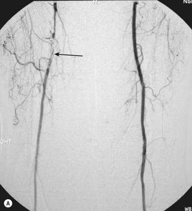
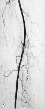
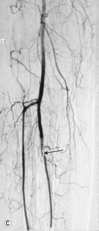
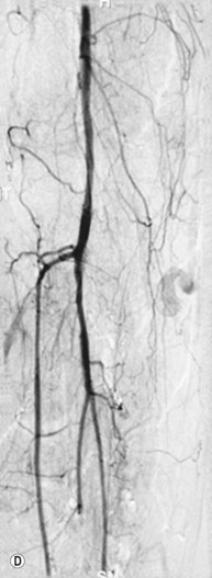
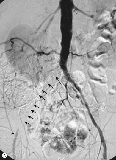
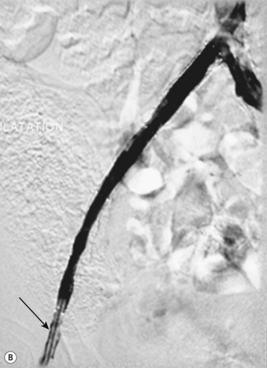
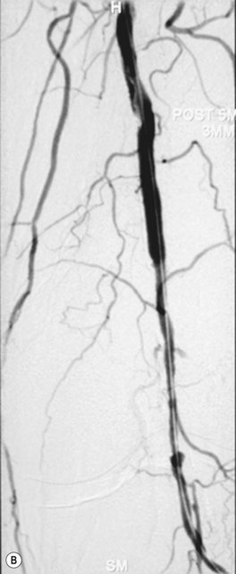
Superficial femoral artery (SFA) angioplasty (Fig. 34.1A–D)


This procedure is most commonly undertaken for the management of critical lower limb ischaemia or short-distance intermittent claudication. Such ischaemia is most likely to be caused by SFA occlusion, rather than a simple stenosis; thus to perform an angioplasty one must first cross the occlusion with a guide wire. This can be difficult, but the use of a hydrophilic guide wire will facilitate successful crossing in the vast majority of cases, with many operators electing to pass the guide wire subintimally. SFA angioplasty is less commonly performed for treatment of intermittent claudication, as generally the results are inferior to those of iliac angioplasty,4,5 and two randomised studies have shown that the results are no better over the long term than those observed after a supervised exercise programme.6,7


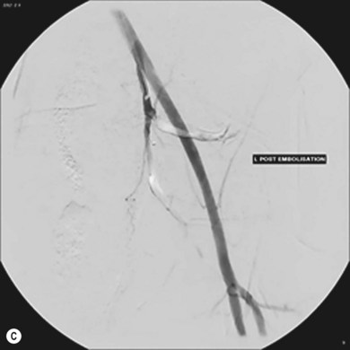
As with iliac angioplasty, SFA lesions can be approached either contralaterally or ipsilaterally. The contralateral approach is the same in technical terms as that used for the iliac vessels. However, the ipsilateral approach to the SFA is technically more difficult, as an antegrade puncture of the CFA is required. To perform an antegrade puncture, the femoral head is first identified under fluoroscopy and its position marked on the skin surface with a metal marker. Local anaesthetic is infiltrated into the skin over the femoral pulse as it is palpated at this level. A puncture needle is introduced first and a guide wire is then introduced along the SFA. It is possible that the guide wire may pass into the profunda femoris, and for this reason it is important to observe its progress under fluoroscopic control. If it proves difficult to enter the SFA, it may be necessary to screen over the needle tip while manipulating it into different positions to facilitate guide wire advancement. When doing this it is very easy for the operator to put their hands into the X-ray beam without realising. The radiographer can prevent or minimise this by centring only on the very tip of the needle, rather than its whole length, and using the collimators appropriately. Antegrade puncture is often used because the distance from the puncture site to the angioplasty site is short, avoiding the need to use very long guide wires. It also avoids any problems associated with catheter manipulation when dealing with tortuous iliac arteries or an acutely angled aortic bifurcation; in the event of a complication occurring, the subsequent management, e.g. aspiration embolectomy, is much more straightforward (Fig. 34.1C,D).


Popliteal artery and the tibial vessels


Lesions in these vessels will only be treated with angioplasty in the presence of critical lower limb ischaemia or short-distance claudication (Fig. 34.2A,B). The potential benefit of angioplasty at these sites in patients with uncomplicated intermittent claudication would be completely outweighed by the potential risk and the likely recurrence rate in the future.8 Technically there is very little difference between angioplasty performed here and elsewhere in the lower limb. Smaller diameter balloons are used, and many operators prefer to use finer guide wires.


Vascular stent insertion


The term ‘stent’ describes a device designed to keep a passage or conduit open. Vascular stents have become accepted as extremely helpful both in maintaining patency where the result of angioplasty alone has been suboptimal, and in certain locations where they provide such markedly superior benefits compared to angioplasty alone that they are considered to be the first line of treatment.


Vascular stents are metallic, commonly made either of stainless steel or nitinol. Nitinol is a nickel–titanium alloy which has great elasticity and ‘shape memory’, which allows it to return to its original state even after significant manipulation and bending. Stents may be self-expanding or balloon expandable. Prior to deployment, stents are compressed onto a delivery catheter; each end of the stent either has radio-opaque markers on the device itself or on the catheter, to facilitate correct positioning. The technique used for deployment of a stent is much the same as that described for angioplasty, with the obvious difference that instead of performing simple balloon dilation, a stent is deployed instead. It will often prove necessary to perform angioplasty prior to stent deployment, and further angioplasty after deployment may be required to ensure that the stent is fully expanded.


Stents are used commonly in the iliac, renal and subclavian arteries. They are being used increasingly in the carotid arteries, although this remains experimental. In the UK, stents have only been used in the SFA as a ‘bail-out’ if angioplasty has resulted in vessel occlusion. However, stents have been used much more freely in the SFA elsewhere, and evidence is starting to show, at least with more modern stent designs, that concerns about low long-term patency rates of stents in the SFA (compared to those of angioplasty alone) may be unfounded. Stents are not used routinely in the popliteal or tibial vessels, though devices are available to be used in the event of a suboptimal result.


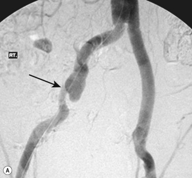
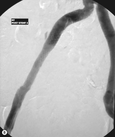
Stenting the iliac artery (Fig. 34.3A,B)


It has been shown that if iliac angioplasty is technically successful there is no advantage in terms of clinical outcome in adding a stent.911 However, in about 50% of cases the outcome from angioplasty is suboptimal, perhaps due, for example, to elastic recoil of the vessel wall or dissection causing flow limitation. Many professionals would add to this and include failure to reduce the intra-arterial pressure gradient across the lesion to less than 10 mmHg as an indication.


The exception to this is the treatment of iliac artery occlusions where, if angioplasty alone is used, there is an incidence of peripheral embolisation of up to 50%.9 For this reason, primary stenting is undertaken when treating iliac occlusions endovascularly. Thus, a self-expanding stent is first deployed across the occlusion and subsequently dilated using an angioplasty balloon.


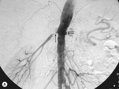
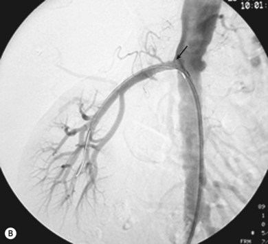
Stenting the renal artery (Fig. 34.4A,B)


Renal artery stenosis is generally caused by one of two pathologies, either fibromuscular hyperplasia or atheroma. Fibromuscular hyperplasia is an uncommon cause of uncontrollable hypertension and responds well to angioplasty alone. Atheromatous renal artery stenosis (ARAS), when it requires treatment, responds very poorly to angioplasty alone, and it has clearly been demonstrated that primary stenting is superior in both the short and the longer term.12 This happens because the vast majority of ARAS occurs at the origin of the vessel and is caused by aortic atheroma rather than true atheroma of the renal artery. Therefore, an expansile force applied to the stenosis causes shear stresses within the aortic plaque, rather than an expansile force within the renal artery lumen. Once the angioplasty balloon is removed the stenosis will frequently recur as the aortic plaque moves back into position.


Balloon-expandable stents are favoured for the treatment of ARAS. In order to avoid the stent being compressed by the aortic plaques, it is necessary to position the stent so that it projects 2–3 mm into the aortic lumen. Such precision is much easier to achieve with balloon-expandable stents, as they do not shorten when they are deployed. Although much improved over older designs, even modern self-expanding stents show some shortening.


Vascular stent grafts


As previously mentioned, stent grafts are used in the treatment of true or false aneurysms. The technology continues to evolve, and it is not possible to say at this point whether stent grafting will replace open surgery in the treatment of aneurysmal disease. However, there is growing evidence to support the use of stent grafts in the treatment of thoracic aortic aneurysms, where the risks of surgery are considerably greater than those of open surgery for abdominal aneurysms.13 Furthermore, there is some evidence to suggest that stent grafting may be of value in patients who would be at greater than average risk for abdominal surgery, for example if they have renal failure.1416 More recently, randomised data have shown a reduction in 30-day mortality when stent grafts are used for abdominal aortic aneurysm repair, compared to open surgery.17,18


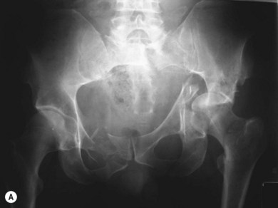
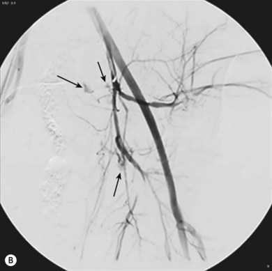
When used for aortic aneurysms, stent graft delivery systems are large and require surgical exposure of one or both common femoral arteries. Smaller aneurysms, such as in the iliac arteries, can be treated without surgical exposure of vessels (Fig. 34.5A,B). Therefore, aortic stent graft procedures are frequently performed in the operating theatre with a mobile image intensifier. A better alternative, which is becoming increasingly available, is to use an angiographic suite that has been constructed to operating theatre standards. This provides a sufficiently sterile environment with a high standard of imaging.


Prior to the stent graft procedure the aneurysm is assessed for the diameter of the proximal and distal landing zones, as well as the overall length of the device. A number of ‘off the shelf’ devices are available, and several manufacturers are able to supply custom-made stent grafts for more complex cases.


Angiographic ‘runs’ are performed to ensure precise positioning of the device. For example, in stent grafting of abdominal aortic aneurysms it is clearly vital to avoid covering (and thereby occluding) the renal arteries with graft material. However, there are devices that have a bare stent at the proximal end which is designed to lie over the renal arteries. Once an image has been selected as the reference image for the deployment of the device it is vital that the C-arm is not moved. Even slight movement can cause errors due to parallax, which could cause misplacement of the stent graft.


Embolisation


Commonly used embolisation agents include gelatin sponge (Fig. 34.6A–C) (for temporary embolisation), polyvinyl alcohol particles and coils (for permanent vessel occlusion). The full range of embolic materials available for clinical use is vast, complex, and includes materials that would require a whole chapter to describe and explain in detail. Embolisation procedures are often complex and time-consuming, and may require the use of superselective coaxial catheter systems; multiple magnified views of the area are needed.


The basic principle of embolisation is to identify the target vessel and place the catheter tip in the correct location prior to introducing the embolic material. Generally one wishes to place the catheter as far distally as possible to avoid embolisation of normal tissue. In addition, when delivering particulate materials it is important to avoid reflux of emboli. It is important, therefore, to use continuous fluoroscopy when injecting such materials.


Some embolisation procedures are relatively simple, such as treatment of varicocoeles. Varicocoeles normally affect the left testis, and occur because the valve at the confluence of the left testicular and the renal vein is incompetent, allowing reflux of blood at systemic venous pressure into the venous drainage of the testis. Treatment involves embolisation of the left testicular vein. The procedure involves placing a catheter in the left renal vein and injecting contrast while screening, and also saving the fluoroscopic image. Once valve incompetence has been confirmed and the anatomy demonstrated, the testicular vein is entered and embolisation coils are placed along its length. Generally patients requiring embolisation of the testicular vein are young, and it is clearly important to minimise radiation dose during this procedure.


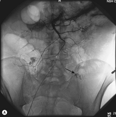
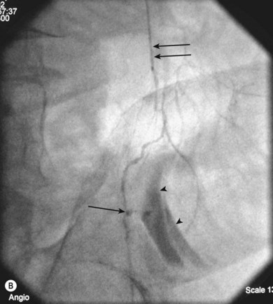
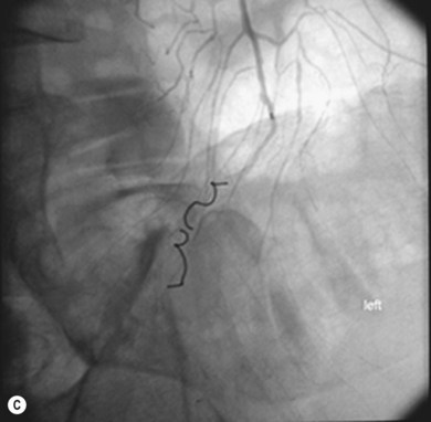
Other procedures are more complex, such as embolisation for gastrointestinal bleeding (Fig. 34.7A–C), and require a more flexible approach to determine the precise anatomy and demonstrate the bleeding point accurately, followed by therapy. Highly complex situations, such as therapy for arteriovenous malformations, may be better referred to centres with a specialist interest in this area.


Venous interventions


Commonly undertaken venous interventions include placement of tunnelled venous lines and insertion of inferior vena cava (IVC) filters. Stents are also used in the venous system; however, the techniques used are very similar to those used in arteries, so it is not necessary to describe them in any greater detail.


Tunnelled central venous lines


Tunnelled central venous lines are used for a variety of purposes, including administration of chemotherapy, total parenteral nutrition and temporary (and occasionally permanent) haemodialysis access. The line is tunnelled subcutaneously; near the point where the tunnel exits the skin it has a Dacron cuff attached to it which becomes incorporated into the tissues, making accidental dislodgement much less likely than with non-tunnelled lines. There are also port systems available in which the entire device can be placed subcutaneously and be accessed percutaneously with a needle for drug administration.


The most commonly accessed vessels are probably the internal jugular veins, followed by the subclavian veins. On occasion, these vessels are occluded; this is particularly the case for patients who have had multiple central lines placed in the past, e.g. for haemodialysis. In these instances it may prove necessary to use alternative vessels, such as the external jugular vein, or even direct puncture of the IVC to provide venous access.


The best method of guiding the vessel puncture is US, which has the clear benefit of avoiding the use of ionising radiation and is recommended by NICE for jugular vein puncture.19 When performing subclavian vein puncture it is possible to opacify the target vein with contrast, and guide the puncture in this way. Fluoroscopy is used to identify the catheter tip when positioning it in the superior vena cava (SVC). The first choice of vein for puncture is the right internal jugular. This vein follows an almost straight course into the right brachiocephalic vein and subsequently the SVC, meaning that there is little potential for kinking of the introducer sheath during insertion. Use of the left internal jugular and subclavian veins is usually straightforward, whereas the use of the right subclavian vein can be difficult, as kinking of the introducer sheath can be a significant problem here.

Only gold members can continue reading. Log In or Register to continue

Stay updated, free articles. Join our Telegram channel

Mar 3, 2016 | Posted by in GENERAL RADIOLOGY | Comments Off on Interventional and therapeutic procedures

Full access? Get Clinical Tree
Get Clinical Tree app for offline access