Intestinal Scleroderma
Michael P. Federle, MD, FACR
Key Facts
Terminology
Progressive systemic sclerosis
Imaging
Multisystemic disorder with immunologic and inflammatory changes
Characterized by atrophy, fibrosis, sclerosis of skin, vessels, and organs
Smooth muscle is replaced by fibrous tissue
Gastrointestinal tract: Most common internal organ system involvement (80-90%)
Esophagus > duodenum > anorectal > small bowel > colon
Small bowel
Marked dilatation of small bowel, especially duodenum and jejunum
Duodenal findings identical to “SMA syndrome”
“Hidebound” small bowel: Atonic with closely spaced thin folds, sacculations
Prolonged transit time with barium retention in duodenum and small bowel up to 24 hours
± pneumatosis intestinalis and pneumoperitoneum
± transient, nonobstructive intussusceptions
Colon
Sacculations on border of transverse and descending colon
Loss of haustrations
Stercoral ulceration (from retained fecal material in rectosigmoid)
Top Differential Diagnoses
SMA syndrome
Sprue-celiac disease
Ileus
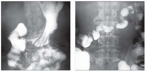 (Left) Film from a small bowel follow through shows dilation of the duodenum with an abrupt narrowing  as it crosses the spine. (Right) Delayed film from the same patient shows barium within the colon, which has a peculiar appearance of sacculations as it crosses the spine. (Right) Delayed film from the same patient shows barium within the colon, which has a peculiar appearance of sacculations  along the mesenteric border. These reflect the muscle atrophy within the bowel wall and its replacement by collagen and fibrosis. along the mesenteric border. These reflect the muscle atrophy within the bowel wall and its replacement by collagen and fibrosis. |
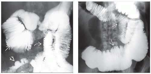 (Left) Small bowel follow through shows a typical dilated duodenum  and a “hidebound” fold pattern of the jejunum with closely spaced, thin transverse folds and a “hidebound” fold pattern of the jejunum with closely spaced, thin transverse folds  , and an atonic, stiff appearance at fluoroscopy. Also note the pseudosacculation , and an atonic, stiff appearance at fluoroscopy. Also note the pseudosacculation  . (Right) 90-minute film from a SBFT shows classic scleroderma of the small bowel with dilated, atonic jejunum and closely spaced, thin transverse folds . (Right) 90-minute film from a SBFT shows classic scleroderma of the small bowel with dilated, atonic jejunum and closely spaced, thin transverse folds  with slow transit. “Pseudo-obstruction” is another descriptive term relevant to this case. with slow transit. “Pseudo-obstruction” is another descriptive term relevant to this case. |
TERMINOLOGY
Synonyms
Progressive systemic sclerosis
Definitions
Multisystem disorder of small vessels and connective tissue of unknown etiology
IMAGING
General Features
Best diagnostic clue
Dilated, atonic small bowel with crowded folds and wide-mouthed sacculations
Other general features
Multisystemic disorder with immunologic and inflammatory changes
Characterized by atrophy, fibrosis, sclerosis of skin, vessels, and organs
Involves skin and parenchyma of multiple organs
GI tract, lungs, heart, kidneys, and nervous system
Gastrointestinal tract (GI) scleroderma
2nd most common manifestation after skin changes (80-90% of patients)
Most common sites: Esophagus > duodenum > anorectal > small bowel > colon
Most frequent cause of chronic intestinal pseudoobstruction
Scleroderma classified into 2 types
Diffuse scleroderma
CREST syndrome (more benign course)
Diffuse scleroderma: Cutaneous and visceral involvement
Severe interstitial pulmonary fibrosis
Organ failure more likely
Associated with antitopoisomerase 1 antibody (anti-Scl 70)
CREST syndrome: Less cutaneous and visceral involvementRelated posts:

Stay updated, free articles. Join our Telegram channel

Full access? Get Clinical Tree


