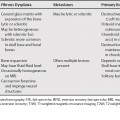
147 Meningiomas and nerve sheath tumors are the most common intraspinal tumors, with meningiomas representing approximately 25 to 40% and nerve sheath tumors representing 16 to 30% of adult intraspinal tumors, with some series reporting nerve sheath tumors as the most frequent intraspinal mass. Because they both commonly arise somewhat laterally in the spinal canal in the intradural extramedullary space (with the nerve sheath tumors most commonly arising from the dorsal sensory roots), they are sometimes difficult to differentiate. Tumors in this space should compress and displace the spinal cord, should expand the cerebrospinal fluid space above and below, and compress the epidural fat. Nerve sheath tumors include neurofibromas and schwannomas, which have been variably called neurinomas, neurolemmomas, and neuromas. Despite several attempts, no technique can reliably distinguish these neoplasms by imaging. Magnetic resonance imaging (MRI) features of nerve sheath tumors reflect their variable histology and growth pattern (Table 147.1).
Intradural Extramedullary Lesions
Nerve Sheath Tumors
| Meningioma | Nerve Sheath Tumor | |
|---|---|---|
| Unenhanced C T | May have calcifications May be hyperdense to spinal cord | May remodel bone/expand neural foramen |
| C T/Myelography | Compress and displace the spinal cord and epidural fat Expand the CSF space above and below* | Compress and displace the spinal cord and epidural fat Expand the CSF space above and below* |
| Unenhanced T1WI | Homogeneous, isointense to mildly hypointense to cord | Heterogeneous, hypointense to isointense, with or without blood products |
| Enhanced T1WI | Homogeneous enhancement | Heterogeneous or peripheral ring enhancement |
T2WI
Stay updated, free articles. Join our Telegram channel
Full access? Get Clinical Tree
 Get Clinical Tree app for offline access

|


