Intradural Schwannoma


Clinical Presentation


The patient is a 32-year-old man with a history of low back and left hip pain that developed 9 months prior and diffuse back pain and back throbbing over the past 3 months. The patient has recently developed lower extremity numbness and burning in the feet and occasionally experiences a sharp radiating pain down the spine (L’Hermitte’s sign). He has been unable to walk any distance and experiences occasional urinary and fecal urgency. He reports one episode of urinary incontinence. Physical examination reveals diminished sensation to pinprick in the lower trunk and below. He has moderately reduced strength in the hip flexors and mildly reduced strength in the feet dorsiflexors. The clinical findings are those of myelopathy with a sensory level in the mid to lower thoracic region.


Imaging Presentation


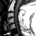
Magnetic resonance (MR) imaging was obtained and revealed a lobulated, dumbbell-shaped, 4 cm in maximal dimension mass located at the T11 level. The mass has a large intradural component and smaller extradural component extending into and enlarging the right T11-12 neural foramen, consistent with nerve sheath tumor (schwannoma or neurofibroma) ( Figs. 65-1 to 65-4 ) .


Figure 65-1


“Dumbbell” Schwannoma, T11 Level.

A large lobulated mass ( arrows ) enhances homogeneously following IV contrast on coronal T1-weighted MRI. The intradural component of the mass is larger, compressing and displacing the spinal cord ( C ) to the left. The extradural component of the mass has enlarged the right T11-12 neural foramen.


Figure 65-2


Illustration of Thoracic Dumbbell Schwannoma.

Similar to that depicted in Figure 65-1 . The schwannoma ( S ) has intradural and extradural components. A large intradural component compresses the spinal cord ( C ) on the right and displaces the cord to the left of midline. The extradural component of the tumor extends out the right T11 neural foramen. P = vertebral pedicle.


Figure 65-3


“Dumbbell” Schwannoma, T11 Level.

Same patient as in Figure 65-1 . Midline sagittal T2 weighted MR image A , T1-weighted image B , and fat-saturated contrast enhanced T1-weighted image C . The mass ( arrow ) is slightly heterogeneous but predominantly isointense relative to the spinal cord on T1- and T2-weighted images. In image C the mass ( arrow ) enhances intensely and homogeneously. T11 = T11 vertebral body.


Figure 65-4


“Dumbbell” Schwannoma, T11 Level.

Same patient as in Figures 65-1 and 65-3 . Parasagittal T2-weighted image A , T1-weighted image B , and fat-saturated contrast enhanced T1-weighted image C . The mass ( arrow ) is slightly heterogeneous on T2-weighted image A . On image T1-weighted image B , the mass ( arrow ) appears more homogeneous and enlarges the right T11-12 neural foramen. On image C , the mass ( arrow ) enhances intensely and homogeneously following IV contrast administration.


Discussion


A schwannoma is a nerve sheath tumor arising from Schwann cells in the nerve sheath. Sometimes referred to as neurilemmoma or neurinoma , a schwannoma is the most common intradural-extramedullary tumor originating within the thecal sac (meningioma is the second most common intradural-extramedullary tumor) ( Fig. 65-5 ) . Approximately 75% of spinal schwannomas arise intradurally; 15% of spinal schwannomas are located in both the intradural and extradural compartments. The majority of other spinal schwannomas are entirely extradural, located in the paraspinal region ( Fig. 65-6 ) . Rarely, intradural schwannomas occur that have an intramedullary component.


Figure 65-5


Small Intradural Schwannoma within Lumbar Thecal Sac.

On post-myelogram axial CT image A in the lumbar region, a small, round, dark filling defect ( arrow ), representing an intradural mass, is located within the contrast filled thecal sac. On corresponding contrast enhanced axial T1-weighted MR image B , the mass ( arrow ) within the thecal sac enhances with contrast and is larger than adjacent normal cauda equina nerve roots. Small intradural schwannomas such as this are often discovered as incidental findings on routine MR imaging studies of the lumbar spine.


Figure 65-6


Left Paraspinal Schwannoma, Upper Thoracic Level.

Axial T2-weighted image A . T1-weighted image B , and fat saturated contrast enhanced T1-weighted image C . The schwannoma ( arrows in image A and B ) is hypointense relative to paraspinal fat on T2-weighted and T1-weighted images. On contrast-enhanced fat-saturated T1-weighted MR image C , the schwannoma enhances intensely and is slightly heterogeneous.


Those schwannomas with intradural and extradural components characteristically have a dumbbell configuration. Small to medium-sized schwannomas are commonly discovered as incidental findings within the thecal sac arising from the cauda equina on routine MR imaging ( Figs. 65-5 , 65-7, and 65-8 ) . Intradural nerve sheath tumors can be lobulated (see Figs. 65-7 and 65-8 ) or spherical in configuration ( Fig. 65-9 ) . So called giant schwannomas may also occur, which are large tumors that can extend for a variable distance into the paraspinal soft tissues ( Fig. 65-10 ) .


Figure 65-7


Lobulated Intradural Schwannoma.

Patient with left S1 radiculopathy. Sagittal unenhanced T1-weighted image A and contrast enhanced T1-weighted image B . The mass ( arrow in image A and B ) enhances homogeneously following IV contrast.


Figure 65-8


Intradural Schwannoma with Extradural Component.

Same patient as in Figure 65-7 . Axial contrast enhanced image A obtained at L5-S1 level, and contrast-enhanced image B obtained at level of S1 lateral recess. A homogeneously enhancing intradural mass ( arrow in image A ) is demonstrated within the thecal sac on the left. The tumor has an extradural component ( large arrow in B ) which compresses and displaces the left S1 nerve root ( S1 ).


Figure 65-9


Intradural Schwannoma Adjacent to Conus Medullaris.

Ovoid intradural mass at L1-2 level simulates a myxopapillary ependymoma. The intradural mass ( arrow ) is hyperintense relative to CSF on sagittal conventional T2-weighted MR image A and on proton density-weighted image B . The mass ( arrow ) is isointense relative to the spinal cord on unenhanced sagittal T1-weighted image C . On sagittal contrast-enhanced image D , the schwannoma ( arrow ) enhances intensely and nearly homogeneously.


Figure 65-10


Giant Paraspinal Schwannoma at C2-3 Level.

Achondroplastic dwarf. Large paraspinal mass demonstrated on unenhanced parasagittal CT image A and axial CT image B . The schwannoma has a slightly hyperdense rim ( long arrows in A and B ) and is relatively hypodense centrally. A component of the mass enlarges the right C1-2 neural foramen ( short arrows in image A ).


Schwannoma is the second most common nerve sheath tumor, the neurofibroma being the most common nerve sheath tumor. Far less common nerve sheath tumors include ganglioneuromas and malignant nerve sheath tumors. Schwannomas arise in the nerve sheath eccentric to the adjacent nerve and are slowly growing tumors. These tumors manifest as isolated masses or as multiple masses anywhere along the spinal axis. Schwannomas are usually isolated lesions except when associated with neurofibromatosis type-2 (NF-2). Most patients with a schwannoma have the sporadic form of the disease; 50% or more of the patients with the more common (sporadic) form have gene mutations that inactivate a portion of chromosome 22 . However, the patients with the sporadic form do not have the other manifestations of neurofibromatosis type 2 (NF-2).


Most schwannomas, solitary or multiple, occur sporadically. The presence of multiple schwannomas does not necessarily mean the patient has NF-2, although multiple schwannomas almost always occur in patients with NF-2. Neurofibromatosis type 2 is an autosomal dominant condition associated with mutations in chromosome 22q (long arm) that occurs in approximately 1 in 30,000 people. Patients with NF-2 usually have multiple schwannomas either intracranially, in the spinal canal, or neural foramina, along with other tumors, including meningiomas and ependymomas, which may occur in the spine or intracranially in NF-2 patients ( Figs. 65-11 to 65-14 ) . The presence of bilateral cranial nerve VIII schwannomas is pathognomonic of NF-2 (see Fig. 65-11 ), but cranial nerve VIII schwannomas may be absent in patients with NF-2. The intradural and paraspinal nerve sheath tumors in NF-2 are usually schwannomas, but some patients with this condition have both nerve sheath schwannomas and neurofibromas. However, the majority of nerve sheath tumors in patients with neurofibromatosis type 1 (NF-1, von Recklinghausen disease) are neurofibromas or plexiform neurofibromas. However, schwannomas can also occur in patients with NF-1.


Figure 65-11


Multiple Intracranial Schwannomas and Meningiomas.

Patient with neurofibromatosis type 2 (NF-2). Axial fat-saturated contrast enhanced T1-weighted MRI obtained through posterior fossa reveals multiple enhancing intracranial schwannomas ( S ) involving cranial nerves V and VIII bilaterally. A meningioma ( M ) is located along the posterior slope of the right petrous bone and another meningioma ( M ) is located to the right of the torcula.

Aug 25, 2019 | Posted by in NEUROLOGICAL IMAGING | Comments Off on Intradural Schwannoma

Full access? Get Clinical Tree

Get Clinical Tree app for offline access