Intrathecal Radiculitis


Clinical Presentation


The patient is a 30-year-old female with severe low back pain and right lower extremity pain in the right S1 distribution. On physical examination, she has diffuse lower extremity weakness, right heel drop when she walks on her toes, and decreased right ankle jerk.


Imaging Presentation


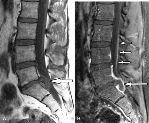
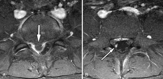
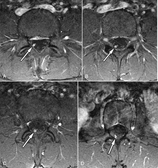
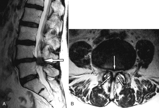
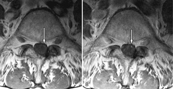
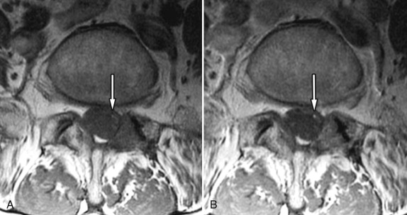
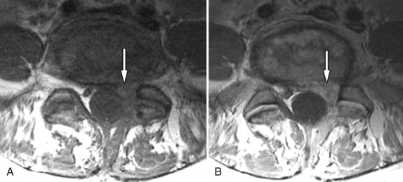
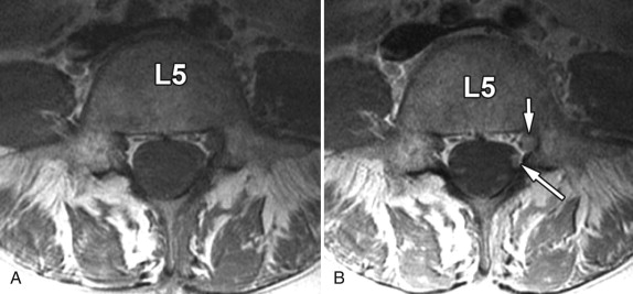
Magnetic resonance (MR) imaging reveals a very large disc extrusion compressing the thecal sac on the right at the L5-S1 level. There is intense contrast enhancement surrounding the disc fragment and diffuse enhancement of the intrathecal right S1 nerve root above the level of the disc herniation, indicating S1 radiculitis ( Figs. 40-1 to 40-3 ) .


Figure 40-1


Disc Extrusion, Nerve Root Inflammation.

Large L5-S1 herniated disc extrusion ( long arrow ) shown on unenhanced T1-weighted MR image A and contrast-enhanced T1-weighted MR image B . There are diffuse enlargement and enhancement of right S1 intrathecal nerve root ( short arrows in image B ). Note intense enhancement along the margin of the T1 hypointense disc fragment on image B representing inflammation.


Figure 40-2


Disc Extrusion, Nerve Root Inflammation.

Axial contiguous contrast-enhanced T1-weighted images at L5-S1 level. Same patient as in Figure 40-1 . In axial image A , the herniated disc ( arrow ) is isointense relative to the intervertebral disc. Intense contrast-enhancement is demonstrated at margin of disc fragment. On axial image B , obtained just superior to the L5-S1 disc herniation at mid-L5 vertebral level, the intrathecal portion of the right S1 nerve root ( arrow ) is enlarged and enhances intensely compared with other nonenhancing intrathecal nerve roots.


Figure 40-3


Disc Extrusion, Nerve Root Inflammation.

Axial images in mid- and upper lumbar regions in same patient as in Figures 40-1 and 40-2 . The right intrathecal S1 nerve root ( arrow in images A through D ) is enlarged and enhances intensely at all visible lumbar levels above the disc herniation.


Discussion


Intrathecal radiculitis is an important pain-producing condition that is frequently not recognized, which is supported by the fact that there exists a paucity of publications in the literature on this subject. Takata and colleagues in 1988 described visible intrathecal nerve root swelling on post-myelogram computed tomography (CT) images in 60% of the patients in their series who had symptomatic lumbar disc herniations. The enlarged intrathecal nerve root is often caused by a disc herniation, central canal stenosis, or postoperative granulation tissue ( Figs. 40-1 to 40-11 ) . The affected nerve corresponds to the patient’s radicular pain distribution. Intrathecal nerve roots of the cauda equina may enhance on MR imaging after IV contrast enhancement if the blood nerve barrier that normally exists is disrupted. The blood-nerve barrier can break down as a result of mechanical factors, trauma, an autoimmune response, or ischemia. Mechanical compression of the nerve root can result in neural edema and eventually ischemic compromise of the nerve root if the condition is prolonged. Although the precise cause of the blood-nerve breakdown is not known, the nerve root enhancement likely reflects a low-grade inflammatory process involving one or more cauda equina nerve roots; hence, we refer to this condition as intrathecal radiculitis . Intrathecal radiculitis may occur preoperatively or postoperatively and may occur with or without nerve root enlargement (see Figs. 40-1 to 40-9 ).


Figure 40-4


Central Canal Stenosis and Left S1 Radiculitis.

Patient with severe L4-5 central canal stenosis and left S1 radiculitis with difficulty walking and left lower extremity pain. Severe central canal stenosis ( arrow ) is demonstrated at the L4-5 level shown on sagittal T2-weighted MR image A . On axial T2-weighted image B , the severe central canal stenosis ( arrows ) is shown to be secondary to disc bulging, ligamentum flavum thickening, and facet hypertrophy.


Figure 40-5


Central Canal Stenosis and Left S1 Radiculitis.

Same patient as in Figure 40-4 . Unenhanced T1-weighted MR image A and contrast-enhanced image B obtained at L4 level. The intrathecal left S1 nerve root ( arrow in A and B ) is not enlarged but enhances following IV contrast at the L4 level, relative to other intrathecal roots.


Figure 40-6


Central Canal Stenosis and Left S1 Radiculitis.

Same patient as in Figures 40-4 and 40-5 . Unenhanced T1-weighted MR image A and contrast-enhanced image B obtained at L3 level. The intrathecal left S1 nerve root ( arrow in A and B ) is not enlarged but enhances following IV contrast at the L3 level, relative to other intrathecal roots. The nerve root enhanced at every lumbar level above the L4-5 stenosis to the level of the conus medullaris ( not shown ).


Figure 40-7


Postoperative Radiculitis.

Postoperative left L5 and S1 radiculopathy secondary to L5 and S1 nerve root inflammation (radiculitis). Unenhanced axial T1-weighted MR image A and contrast-enhanced axial image B . Relatively T1 hypointense scar/granulation tissue ( arrow in image A ) located anterolateral to thecal sac at the L5-S1 level. The postoperative scar/granulation tissue ( arrow in image B ) enhances following IV contrast.


Figure 40-8


Postoperative Radiculitis.

Postoperative left S1 and L5 radiculitis. Same patient as in Figure 40-7 . L5 vertebral level. Intrathecal portions of the nerve roots are not well seen on axial unenhanced T1-weighted MR image A . On corresponding axial contrast-enhanced T1-weighted MR image B , the left S1 root ( long arrow ) is enlarged and enhances. Extrathecal portion of the L5 nerve root ( short arrow ), located in the left L5 lateral recess, is also enlarged and enhances to a greater degree compared to corresponding normal L5 nerve root on the right.

Only gold members can continue reading. Log In or Register to continue

Stay updated, free articles. Join our Telegram channel

Aug 25, 2019 | Posted by in NEUROLOGICAL IMAGING | Comments Off on Intrathecal Radiculitis

Full access? Get Clinical Tree

Get Clinical Tree app for offline access