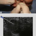
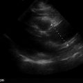
Point-of-care ultrasound (POCUS) has been used clinically since the 1980s. Improved machine image quality, portability, lower cost, and increased evidence for the diagnostic utility of point-of-care ultrasound has moved the probe firmly into the hand of the bedside practitioner. In the time-pressured setting of the emergency department (ED), clinicians have discovered new uses and created innovative workflows to integrate bedside ultrasound into clinical decision making. Critical care and other fields have followed suit, and burgeoning activity within general internal medicine is creating a niche for this imaging method. We clearly believe that point-of-care ultrasound benefits patients and physicians by improving diagnostic accuracy while bringing the physician back to the bedside and protecting patients from ionizing radiation. After your first time finding the unexpected cardiac tamponade in a patient with undifferentiated shock or getting a peripheral IV in the otherwise “impossible” patient, we think you will see the value in point-of-care ultrasound, as well.
New users of ultrasound are tempted to immediately integrate images into their clinical decision making. In medicine, however, bad information is worse than no information, and our conceptual framework emphasizes the following skills that need to be acquired by new users: 1) know the indications for a particular point-of-care ultrasound examination. 2) know how to acquire the images needed for high-fidelity. 3) know how to interpret the information acquired, 4) know how to clinically integrate the findings.
Our experience running an emergency medicine ultrasound fellowship since 2004 and creating the first combined emergency medicine/internal medicine (EM/IM) ultrasound fellowship in 2016, drove us to write this book. Our team includes senior EM doctors, IM doctors, and sonologist educators with more than 50 cumulative years of experience using and teaching ultrasound. Although numerous high-quality textbooks on point-of-care ultrasound have been written by luminaries in the field, it is impractical to bring a large text to the patient’s bedside. And while there are several app- and Web-based resources, there is nothing like paper in hand—paper never runs out of battery or drops the Internet connection. We realize that trainees often need just a small nudge at the point of care: a reminder of what images to acquire and how to get them, or a quick glance at a pathologic image in comparison with a normal one. This book fills that need, providing those point-of-care tips for when a learner can’t have an expert at their shoulder. It doesn’t replace the comprehensive ultrasound text, but for the learner at all levels, this pocket guide will alleviate the anxiety of scanning while preventing some of the more common pitfalls.
Each chapter in this book is divided into four sections: Key Images, Acquisition Tips, Interpretation and Pitfalls, and Examples of Pathology. They are conveniently located on cards that can be popped out and brought with you to the bedside as a reference. Take notes on the cards! Check off the scans you’ve done! If your favorite scan isn’t included, fear not: We are planning to release multiple click-in modules for different scans and different specialties. Many of the images in the book are actually stills from video files that are available as a part of the purchase at www.accessmedicine.com/POCUS. We hope that this book will allow you to feel confident as you learn ultrasound at the same time as ensuring that your patients are receiving the best possible care.
Point-of-care ultrasound is indicated for:
Performing most bedside procedures for which a needle or device would otherwise be placed using landmarks and physical exam (e.g., thoracentesis, paracentesis, central line)
Time-sensitive conditions (e.g., cardiac tamponade, pneumothorax)
Diagnostic uncertainty (e.g., urinary retention, questionable pleural effusion on chest x-ray)
To narrow an otherwise broad differential diagnosis (e.g., new chest pain, dyspnea, shock)
To assess the effectiveness of an intervention (e.g., reassessment of the Inferior Vena Cava (IVC) after a fluid bolus, B-line assessment of the lungs after diuretics)
Stay updated, free articles. Join our Telegram channel

Full access? Get Clinical Tree


