An Approach to Image Interpretation
The first step in medical imaging is to examine the patient and determine the possible cause of his or her problem. Only after this is done can you decide which imaging study is the most appropriate. A vast number of algorithms and guidelines have been developed, but no definite consensus exists on the “right” one for a given symptom or disease because a number of imaging modalities have similar sensitivities and specificities. In this text I provide tables of appropriate initial imaging studies for various clinical situations. When possible, these tables are based on the published literature and recommendations of professional societies. When this is not possible, I give you my opinion based on 45 years of clinical practice.
What should you expect from an imaging examination? Typically one expects to find the exact location of a problem and hopes to make the diagnosis. Although some diseases present a characteristic picture, most can appear in a variety of forms, depending on the stage. As a result, image interpretation will yield a differential diagnosis that must be placed in the context of the clinical findings.
Examination of images requires a logical approach. First you must understand the type of image, the orientation, and the limitations of the technique used. For example, I begin by mentally stating, “I am looking at a coronal computed tomography (CT) scan of the head done with intravenous contrast.” This is important, because intravenous contrast can be confused with fresh blood in the brain.
Next I look at the name and age on the image label to avoid mixing up patients, and this allows making a differential diagnosis that applies to a patient of that age and sex. You would not believe the number of times that this seemingly minor step will keep you from making dumb mistakes.
The next step is to determine the abnormal findings on the image. This means that you need to know the normal anatomy and variants of that particular part of the body, as well as their appearance on the imaging technique used. After this, you should describe the abnormal areas, because it will help you mentally order a differential diagnosis. The most common mistake is to look at an abnormal image and immediately name a disease. When you do this, you will find your mind locked on that diagnosis (often the wrong one). It is better to say to yourself something like, “I am going to give a differential diagnosis of generalized cardiac enlargement with normal pulmonary vasculature in a 40-year-old man,” rather than to blurt out “viral cardiomyopathy” in a patient who really has a malignant pericardial effusion.
After practicing for 20 years or so, a radiologist knows the spots where pathology most commonly is visualized. Throughout this text, I point out the high-yield areas for the different examinations. Although no absolute rules exist, knowing the pathology and natural history of different diseases will help you. For example, colon cancer typically metastasizes first to the liver rather than the lungs, whereas sarcomas preferentially metastasize to the lungs rather than the liver.
After reviewing the common causes of the imaging findings that you have observed, you should reorder the causes in light of the clinical findings. At this point, you probably think that you are finished. Not so. Often a plethora of information is contained in the patient’s image files or in the hospital’s computer information system. This comes in the form of previous findings and histories supplied for the patient’s other imaging examinations. Reviewing the old reports has directed me to areas of pathology on the current image that I would have missed if I had not looked into the medical information system. A simple example is a pneumonia that has almost but not completely resolved or a pulmonary nodule that, because of inspiratory difference, is hiding behind a rib on the current examination.
You probably think that you are finished now. Wrong again. A certain number of entities could cause the findings on the image, but you just have not thought of them all. After I have finished looking at a case, I try to go through a set sequence of categories in search of other differential possibilities. The categories I use are congenital, physical/chemical, infectious, neoplastic, metabolic, circulatory, and miscellaneous.
X-Ray
Regular x-rays (plain x-rays, also sometimes called radiographs ) account for about 75% of imaging examinations. X-ray examinations, or plain x-rays, are made by an x-ray beam passing through the patient. The x-rays are absorbed in different amounts by the various tissues or materials in the body. Most of the beam is absorbed or scattered. This represents deposition of energy in the tissue but does not cause the patient to become radioactive or to emit radiation. A small percentage of the incident radiation beam exits the patient and strikes a detector.
The historical imaging receptor was a film/screen combination. The x-ray beam would strike a fluorescent screen, which produced light that exposed the film, and then the film was developed. Newer systems are called computed radiography or digital radiography . In computed radiography, the x-rays strike a plate that absorbs the x-rays and stores the energy at a specific location. The plate is then scanned by a laser, which releases a point of light from the plate. The location is detected and stored in a computer. In digital radiography detector systems, the x-ray hits a detector and then is converted to light or an electrical charge immediately. Once either type of image is stored in the computer, it can be displayed on a monitor for interpretation or transmitted to remote locations for viewing.
Four basic tissue densities, or shades, are visible on plain x-rays. These are air, fat, water (blood and soft tissue), and bone. Air is black or very dark. On regular x-rays and CT scans, fat is generally gray and darker than muscle or blood ( Fig. 1.1 ). Bone and calcium appear almost white. Items that contain metal (such as prosthetic hips) and contrast agents also appear white. The contrast agents generally used are barium for most gastrointestinal studies and iodine for most intravenously administered agents.

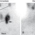
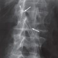
Remember that standard or plain x-rays are two-dimensional presentations of three-dimensional information. That is why frontal and lateral views are often needed. Without these, mistakes can easily be made. You must remember that an object visualized on a specific view is somewhere in the path of the x-ray beam (not necessarily in the patient). If an object projects outside the patient on any view, it is outside the patient. However, even if an object projects within the patient on two orthogonal views, it can still be located outside the patient ( Figs. 1.2 and 1.3 ). Each additional view needed to make a diagnosis requires an additional x-ray exposure and therefore adds to the patient’s radiation dose. Radiation doses from various examinations are given in the Appendix.


The terminology used to describe images is usually quite straightforward. Chest and abdominal radiographs are referred to as upright or supine, depending on the position of the patient. In addition, chest x-rays are usually described as posteroanterior (PA) or anteroposterior (AP) ( Fig. 1.4 ). These terms indicate the direction in which the x-ray beam traversed the patient on its way to the detector. PA means that the x-ray beam entered the posterior aspect of the patient and exited anteriorly. AP means that the beam direction through the patient was anterior to posterior. A left lateral decubitus view is one taken with the patient’s left side down.

Position is important to note, because it can affect magnification, organ position, and blood flow and therefore significantly affect image interpretation. For example, the heart appears larger on AP than on PA images because on an AP projection the heart is farther from the detector and is magnified more by the diverging x-ray beam. It also appears larger on supine than on upright images because the hemidiaphragms are pushed up, making the heart appear wider. Portable chest images are taken not only in the AP projection but also with the tube closer to the patient than on standard upright images. This magnifies the heart even more.
Use of contrast agents permits visualization of anatomic structures that are not normally seen. For example, intravenously or intra-arterially injected agents allow visualization of blood vessels ( Fig. 1.5 ). If imaging is done with standard format, the blood vessels appear white. Digital imaging allows subtraction or removal of unwanted structures, such as the bones, from an image (see Fig. 1.5B ). Often the computer manipulation is done in such a way that the arteries may appear black instead of white, although this usually does not present a problem in interpretation.

Contrast agents are used to fill either a hollow viscus (such as the stomach) or anatomic tubular structures that can be accessed in some way (such as blood vessels, ureter, and common bile duct). When you see an abnormality on one of these studies, you must determine whether the location is intraluminal, mural, or extrinsic. This usually requires seeing the abnormality in perpendicular views ( Fig. 1.6 ). Unless you are careful about this determination, you will make errors in diagnosis.

Related posts:


Stay updated, free articles. Join our Telegram channel

Full access? Get Clinical Tree


