Introduction to PET/CT Imaging
Todd M. Blodgett, MD
Alex Ryan, MD
Marios Papachristou, MD
Introduction to PET/CT Imaging
PET Biochemistry
FDG is taken up by many malignancies due to overexpression of glut-1
Some malignancies may not have significant upregulation of glut-1 and may not be visualized well on FDG PET
PET Physics
PET imaging uses positron emitting radioisotopes such as F-18
F-18 is attached to glucose, resulting in FDG
FDG is taken up by cells, preferentially by many malignant cells
FDG is a marker of cellular metabolism but is not itself metabolized
AC Algorithms
CT is used for attenuation correction with PET/CT scanners
Obviates the need for a separate transmission scan leading to overall decease in scan times
Typical Scan Protocol
Patients are injected with FDG and remain in a quiet room for 1-2 hours
Patients scanned CT first then PET
PET/CT Increasingly Important in
Diagnosis, staging, and restaging of cancer
Image-guided radiation therapy planning
Treatment monitoring
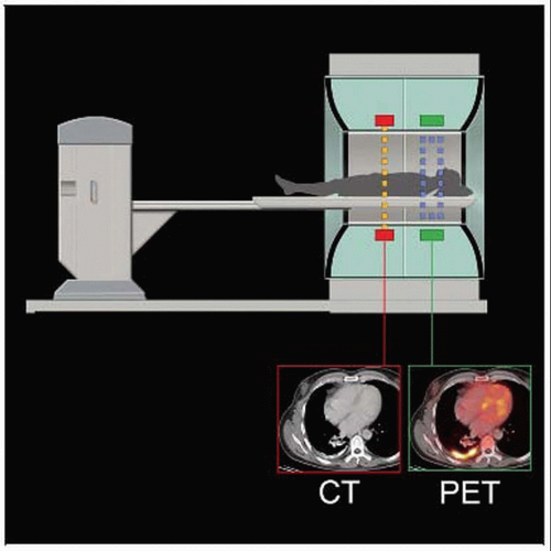 Graphic depicts a typical PET/CT scanner, which houses both CT and PET scanners in a single unit. |
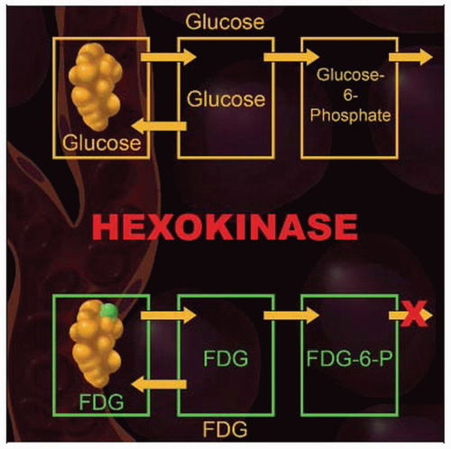 Graphic shows the metabolic pathway for FDG. It enters the cell, undergoes the first step of cellular metabolism, but is not further metabolized. FDG is trapped in the cell long enough to appear on images of the patient. |
TERMINOLOGY
Abbreviations
Fluorodeoxyglucose (FDG), standard uptake value (SUV), Hounsfield unit (HU), attenuation correction (AC), fine-needle aspiration (FNA)
Positive predictive value (PPV), negative predictive value (NPV)
IMAGING ANATOMY
General Anatomic Considerations
Hardware approach to image fusion allows accurate registration of anatomic and metabolic images
PET and CT scanners are housed in single device
Single gantry passes patient through both scanners without interim repositioning
Motion between the CT and PET portions of a PET/CT scan will cause significant misregistration
PET BIOCHEMISTRY
FDG Uptake
Enhanced glycolysis in many malignant cells leads to increased FDG uptake
Glucose transmembrane transporter glut-1 is overexpressed
Not all malignant cells overexpress glut-1 transporter
These malignancies may not take up significantly elevated levels of glucose or FDG
FDG enters the cell and is a substrate for hexokinase, the first enzyme of glycolysis
Hexokinase phosphorylates FDG to FDG-6-phosphate
Metabolic activity of FDG ceases at that point and FDG remains trapped in the cell long enough to image the patient
PET PHYSICS
Hardware
History
First PET/CT scanner became operational in 1998, and first commercial scanners appeared in 2001
Current state of the art incorporates multi detector row CT with high resolution PET scanners
Larger patient ports (70 cm or larger)
Aids in radiation planning and accommodates increasing dimensions of average patient in USA
Technical considerations
Smaller detectors contribute to improved resolution
For example, 4×4 mm lutetium oxyorthosilicate detectors offer slightly higher PET resolution than 6×6 mm detectors
Gadolinium oxyorthosilicate and lutetium oxyorthosilicate scintillators
Result in lower rates of both scattered photons and random coincidences compared with bismuth germinate scintillators
Generally offers improved whole-body 3D imaging
Radiotracers
Radioisotope most commonly used in PET imaging is fluorine-18Related posts:

Stay updated, free articles. Join our Telegram channel

Full access? Get Clinical Tree


