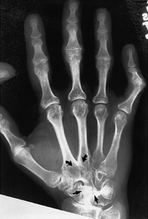
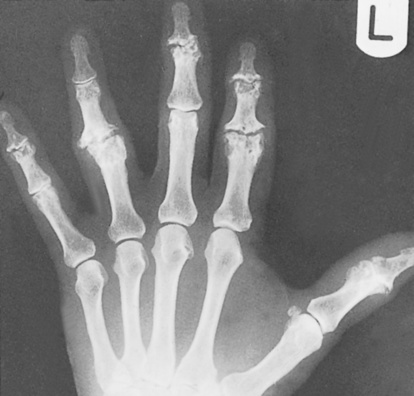
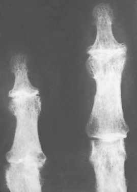
• This is the most common joint disorder – it is a balance between degenerative joint destruction (stressed bone) and repair (non-stressed bone) • Primary OA: there is no underlying cause • Secondary OA: joints are damaged by previous disease Central erosions and marginal osteophytes (a ‘gull wing’ pattern) affecting the DIP joints • Differential: psoriasis and rheumatoid arthritis – however these will have no 1st CMC joint involvement and will have marginal (as opposed to central) erosions with no true osteophyte formation • There is recurrent bleeding into a joint due to a deficiency of blood clotting factors • Acute: joint effusion and oedema • Chronic: juxta-articular osteoporosis resulting from haemorrhage and periarticular hyperaemia Conditions associated with neuropathic arthropathy* • This is otherwise known as juvenile rheumatoid arthritis or juvenile chronic arthritis • It is an inflammatory disorder of the connective tissues characterized by joint swelling, pain and tenderness affecting ≥ 1 joints for at least 6 weeks in patients who are < 16 years of age • Acute systemic onset type (Still’s disease): constitutional symptoms • Oligoarthritis: ≤ 4 joints involved during the 1st 6 months, usually progressing to: • Polyarthritis: ≥ 5 joints involved (both presentations are equally common) • Systemic arthritis: arthritis + a systemic illness • A multifocal entity characterized by ‘flowing’ ligamentous spinal ossification involving • There is no apophyseal or sacroiliac joint fusion (cf. ankylosing spondylitis)
Joint disease
INTRODUCTION
CLINICAL AND RADIOLOGICAL FINDINGS IN JOINT DISEASE
Condition
Site of involvement
Discriminatory findings
Primary osteoarthritis (F>M  > 45 years)
Hands
Large joints (e.g. hip, knee)
Spine
PIP and DIP joint involvement (Heberden’s and Bouchard’s nodes)  no osteopenia
Joint space narrowing  subchondral sclerosis
  subchondral cysts
  marginal osteophytes
Degenerative disc disease  spondylosis deformans
  apophyseal joint involvement
  spinal stenosis
  foraminal stenosis
Erosive osteoarthritis (affects middle-aged females)
Hands
PIP and DIP joint involvement  joint ankylosis
  ‘gull-wing’ deformities (central erosions and marginal osteophytes)
Rheumatoid arthritis (F>M  Rh factor positive)
Hand and wrist
Large joints
Spine
Symmetrical arthritis  MCP and PIP joint involvement
  periarticular (early) and diffuse (late) osteopenia
  marginal erosions
  subluxation (swan neck and boutonnière deformities)
  periostitis is uncommon
Joint space narrowing  marginal erosions
  synovial cysts
  protrusio acetabulae
Atlantoaxial subluxation
Juvenile idiopathic arthritis (M = F  affects children)
Hands
Large joints (e.g. knee)
Cervical spine
Joint ankylosis  florid periosteal reaction
  osteopenia
Abnormalities of growth and maturation  epiphyseal overgrowth and premature closure of the physis
  widened intercondylar notch
Apophyseal joint fusion  atlantoaxial subluxation
Psoriatic arthritis (M>F  nail changes
  HLA-B27 +ve)
Upper extremities (e.g. hands and feet)
SI joints
Spine
‘Sausage’ digit  DIP joint involvement
  terminal tuft erosion
  pencil-in-cup deformity
  joint ankylosis
  arthritis mutilans
  periosteal reaction
  no osteopenia
Asymmetric or unilateral sacroiliitis
Coarse syndesmophytes
Reiter’s syndrome (affects young male adults)
Lower extremities (e.g. foot)
Spine
SI joints
Hallux involvement  periosteal reaction
  calcaneal erosions
  osteopenia not prominent
Coarse syndesmophytes
Asymmetric or unilateral sacroiliitis
Ankylosing spondylitis (M>F  affects young adults
  HLA-B27 +ve in 95%)
SI joints
Spine
Pelvis
Bilateral symmetrical sacroiliitis  ankylosis
Anterior vertebral body squaring  syndesmophytes
  paravertebral ossification
  bamboo spine
‘Whiskering’ of the iliac crests and ischial tuberosities
Enteropathic arthropathies
SI joints
Symmetrical sacroiliitis
Gout (M>F)
Hands and feet (especially the great toe)
MTP joint of the great toe  juxta-articular erosions
  punched-out lesions with an overhanging margin
  no periarticular osteopenia
  tophi
tophi
CPPD crystal deposition disease (M = F)
Any peripheral joint  predilection for the knee
Degenerative changes  chondrocalcinosis
  paucity of subchondral sclerosis
HA crystal deposition disease (M = F)
Predilection for the shoulder (supraspinatus tendon)
Periarticular calcification
Haemochromatosis (M>F)
Hands
2nd and 3rd MCP joint involvement (‘squared’ metacarpal heads)  joint space narrowing
  ‘hook-like’ osteophytes
  numerous subchondral cysts
Alkaptonuria (ochronosis) (M = F)
Intervertebral discs  SI joints
  large joints
Degenerative changes: disc calcification  joint space narrowing
  periarticular sclerosis
Systemic lupus erythematosus (F>M  affects young adults)
Hands
Reversible MCP joint subluxation
Scleroderma (F>M  affects adults)
Hands
IP joint arthritis  acro-osteolysis
  soft tissue calcifications
Mixed connective tissue disease (overlap syndrome)
Hands
PIP joint, MCP joint, mid-carpal involvement  soft tissue swelling, calcifications or atrophy
Multicentric reticulohistiocytosis (F>M)
Hands and feet
DIP joint and carpal involvement  soft tissue swelling
  articular erosions
  no osteopenia
Polymyositis / dermatomyositis
Proximal extremities
Hands
Soft tissue calcification
DIP joint erosions
Sarcoidosis
Distal and middle phalanges of the hands and feet
Punched-out cyst-like lesions  ‘lace-like’ appearance
Haemophilic arthropathy (affecting males – but with female carriers)
Predilection for large joints (e.g. knee)
Epiphyseal overgrowth  juxta-articular osteopenia
  erosion and cartilage destruction
  widened intercondylar and trochlear notches
  squared patella
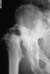
Neuropathic arthropathy
Any joint
5 ‘D’s’: normal bone Density  joint Distension
  bony Debris
  joint Disorganization
  Dislocation
Hypertrophic osteoarthropathy
Tubular bones (radius and ulna > tibia and fibula)
Diaphyseal and metaphyseal painful periostitis
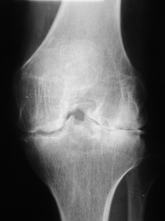
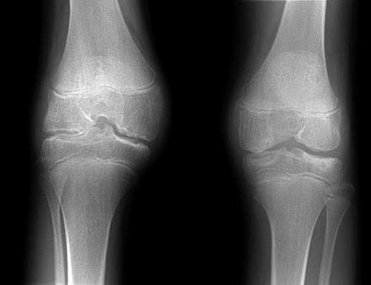
OSTEOARTHRITIS
DEFINITION
 it occurs in the context of normal biomechanical forces
 The joints most at risk: thumb base
  DIP joints
  acromioclavicular joints
  knees
knees  hips
hips  1st MTP joints
  spinal apophyseal joints
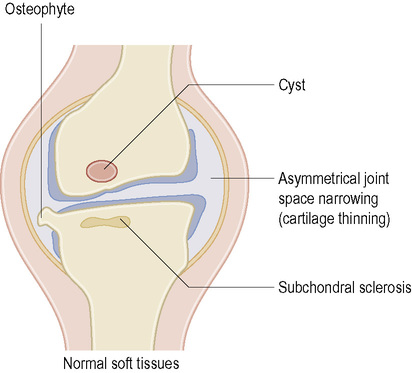
PEARLS
XR
Radiological finding
Pathological cause
Localized joint space narrowing
Articular cartilage fibrillation, ulceration and erosion lead to changes in collagen and protein polysaccharide structure of cartilage. This results in reduced turgor
Subchondral bony sclerosis
Increased osteoblastic activity resulting in new bone formation and increased cellularity of the subchondral bone
Osteophyte formation (most commonly marginal)
Cartilage and bone proliferation and revascularization of remaining cartilage
Bone cysts and bone collapse
Subchondral micro-fractures and passage of synovial fluid under pressure through the damaged cartilage to excavate a subchondral cyst
Gross deformity with subluxation
Ligamentous laxity resulting from mechanical force applied after the distortion of capsular structures
Loose bodies
Fragments of bone and cartilage become separated and, if not resorbed, become loose in the joint. They may reattach to the membrane, become vascularized and undergo endochondral ossification
Fibrocartilage or hyaline cartilage calcification
This is usually due to calcium pyrophosphate deposition disease (CPPD). The reparative response is usually quite florid
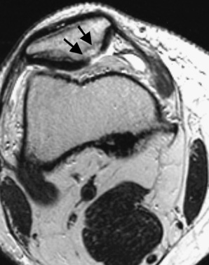
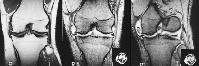
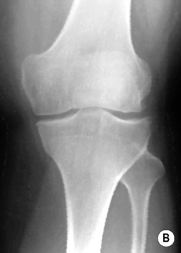
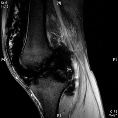
HAEMOPHILIC AND NEUROPATHIC ARTHROPATHIES
HAEMOPHILIC ARTHROPATHY
DEFINITION
 the repeated intra-articular bleeding leads to villous synovial hypertrophy with the accumulation of haemosiderin within macrophages
RADIOLOGICAL FEATURES
Location
XR
 increased radio-opacity of the periarticular soft tissues and synovium (due to haemosiderin deposition)
  articular erosion and cartilage destruction (due to a thickened synovium)
  secondary osteoarthritis (subchondral cysts are common)
 Knee: a widened intercondylar notch
  a squared patella
  it demonstrates similar appearances to juvenile rheumatoid arthritis
 Stage I: soft tissue swelling (± joint effusion)
  normal joint surfaces
 Stage II: stage I + periarticular osteoporosis
  epiphyseal overgrowth
 Stage III: erosions, sclerosis and subchondral cysts
  the joint space is preserved
 Stage IV: stage III + focal or diffuse joint space narrowing
 Stage V: a stiff contracted joint with significant degenerative change
Condition
Prevalence of arthropathy
Joints most commonly affected
Congenital insensitivity to pain
100%
Ankle, tarsal, knee, hip
Syringomyelia
20–50%
Shoulder, elbow, wrist, cervical spine
Neurosyphilis
5–10%
Knee, hip
Diabetes mellitus
1%
Midfoot, forefoot
Alcohol related
Rare
Foot
JUVENILE IDIOPATHIC ARTHRITIS AND DISH
JUVENILE IDIOPATHIC ARTHRITIS (JIA)
DEFINITION
CLINICAL PRESENTATION
 hepatosplenomegaly
  there is little joint involvement
 there is often asymmetrical involvement of the peripheral joints
 there can also be a psoriatic arthritis and an enthesitis-related arthritis
DIFFUSE IDIOPATHIC SKELETAL HYPEROSTOSIS (DISH) (FORESTIER’S DISEASE)
DEFINITION
 4 contiguous vertebrae with preservation of the underlying disc height (cf. degenerative disc disease)
 there is hyperostosis of certain ligamentous attachments
Joint disease

 systemic, metabolic or endocrine disorders (e.g. rheumatoid arthritis, ochronosis, haemochromatosis)
  crystal deposition disease
  neuropathic disorders
  congenital hip dislocation
  bone dysplasias
 reduced movement
  joint crepitus (± effusion)
  early morning stiffness
 primary and secondary OA have similar radiological appearances
 subchondral cysts and sclerosis
  marginal osteophytes
  loose bodies
  chondocalcinosis
 osteoporosis and ankylosis are not features
 distal and proximal IP joint prominences are commonly seen (Heberden’s and Bouchard’s nodes, respectively)
 the medial joint compartment shows the greatest narrowing (as it is subject to greater stressors)
  it usually affects the lateral facet of patellofemoral joint
  there is articular and meniscal cartilage chondrocalcinosis
 there is a tendency for superolateral femoral head subluxation (but there may be lateral restraining osteophytes)
  rarely medial migration can lead to protrusio acetabuli
 destructive changes outstrip productive changes and can mimic an erosive arthritis (such as psoriatic arthropathy)
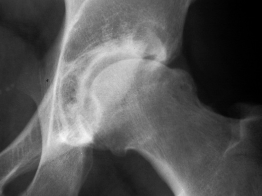

 factor VIII deficiency
 factor IX deficiency
 joint deformities
 small peripheral joints are rarely involved
 there is neither uniform nor symmetrical joint involvement
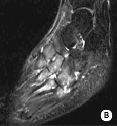
 it can differentiate between acute and chronic soft tissue bleeding
 it occurs particularly within the femur and around the pelvis
  it presents as a painless expanding mass, with soft tissue and bone destruction
 T2WI: central high SI (due to blood breakdown products)
 resorption of the ends of affected bones results in sharp pointed ends
  there is an absence of osteoporosis, sclerosis, fragmentation or soft tissue debris
  it may lead to joint dislocation
 it begins with a joint effusion
  the joint spaces are initially widened but then narrowed
  there is marked bony sclerosis (and no osteoporosis)
  fragmentation of the articular surfaces results in bony debris, which may later fuse into a large dense and well-organized corticated bony mass (± fusion with the underlying bone or dissection into the muscle planes)
  there is periosteal new bone formation
  subluxation and dislocation preceeds total joint disorganization
  pathological fractures can occur
 joint Distension
  bony Debris
  joint Disorganization
  Dislocation
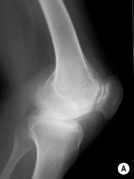
 also involved: feet, shoulders, elbows and hips
 synovitis
  synovial hyperplasia and pannus formation
  a widened joint space (secondary to an effusion)
  periarticular osteopenia
 cartilage loss
  there is often extensive ankylosis (affecting the CMC and mid-carpal joints)
  joint space narrowing
 a widened intercondylar notch of the knee
  overtubulated diaphyses
  enlarged and osteoporotic epiphyses with compression fractures
  florid periosteal new bone formation within the phalanges, metacarpals and metatarsals (cf. RA)
  enlarged, irregular and squared carpal bones (due to erosion and repair)
 atlantoaxial subluxation is common
  compression fractures
  scoliosis (with advanced disease)
 contractures
  AVN (due to JIA or the resultant steroid therapy)
 back pain and stiffness
  tendinosis (commonly affecting the elbow and heel)
 ossification of the anterior longitudinal ligament is seen at the affected level (up to 2cm)
 superior ⅓ of the sacroiliac joints
  symphysis pubis
  calcaneus
  tarsal bones
  patella
  olecranon
  humerus
  hands
hands