Joint Injection: Lower Limb
KEY FACTS
Procedure
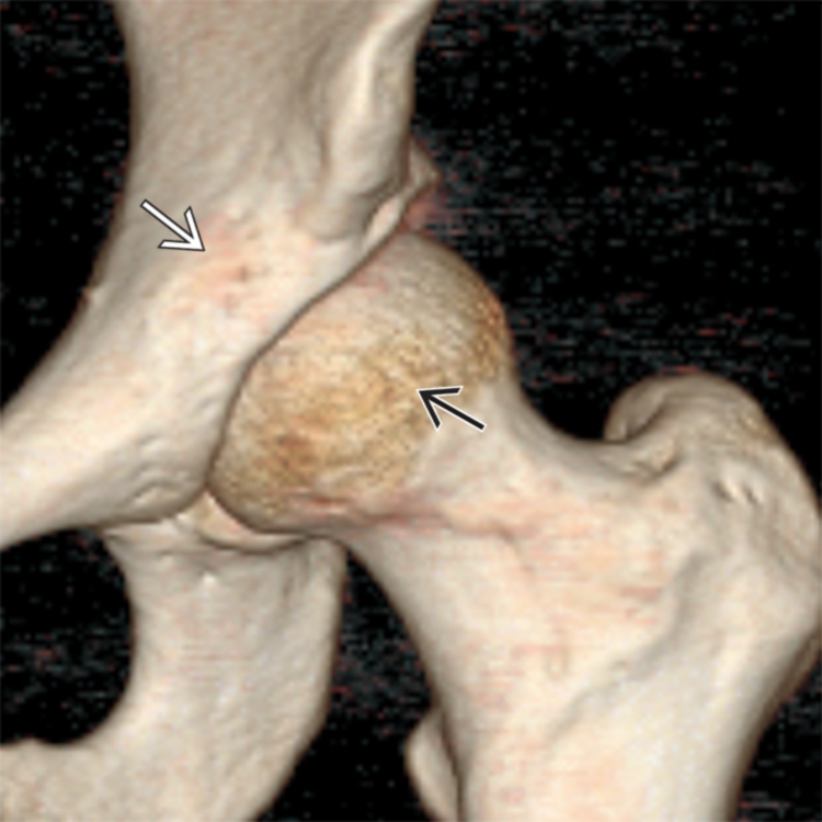
CT volume rendering shows bony anatomy on anterior aspect of the hip joint between the acetabulum  and the femoral head
  .
.
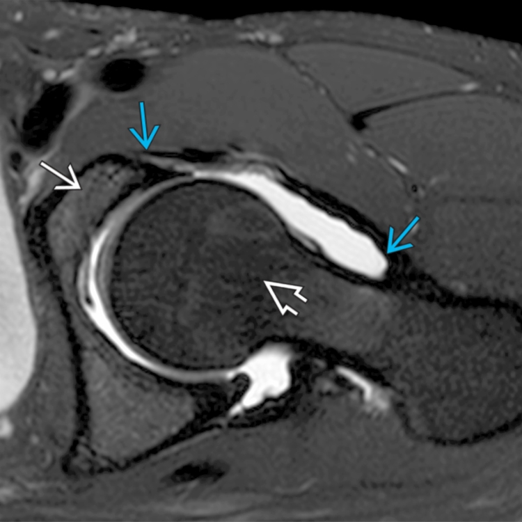
Axial T2 FS MR shows the hip joint between the acetabulum  and femoral head
  . The attachments of the anterior capsule to the lip of the acetabulum and to the base of the femoral neck are shown
  .
.
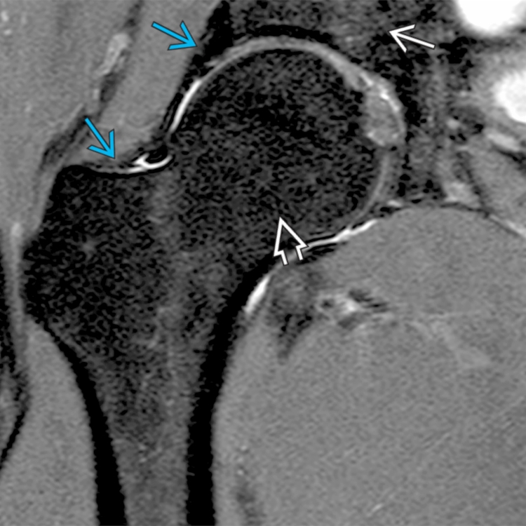
Coronal T2 FS MR shows hip joint between acetabulum  and femoral head
  . The attachments of the anterior capsule to the lip of the acetabulum and the base of the femoral neck are shown
  .
.
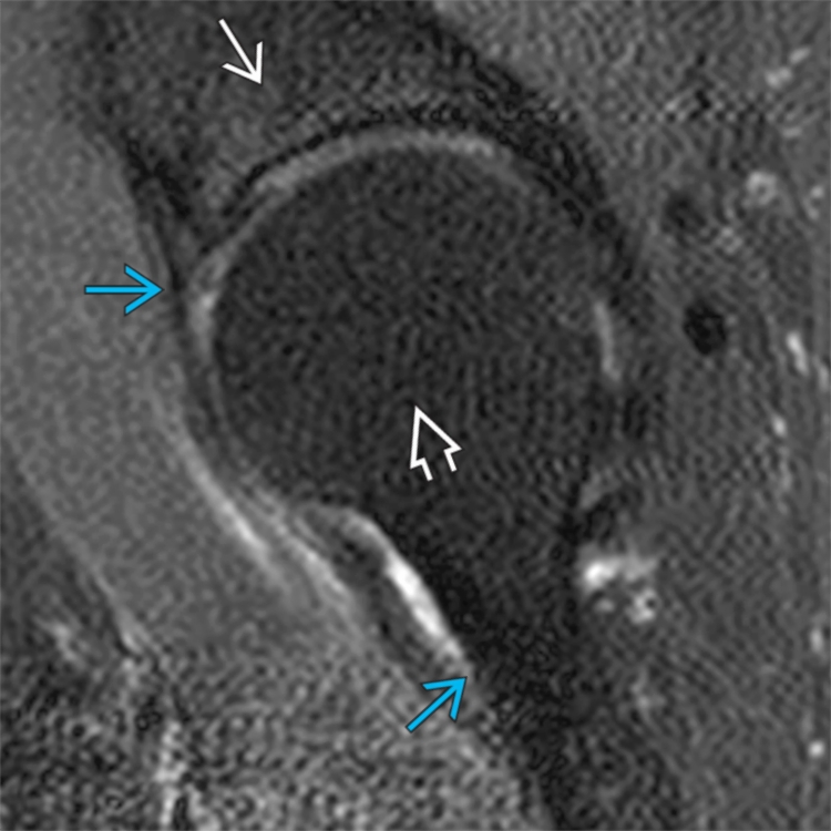
Sagittal T2 FS MR shows hip joint between acetabulum  and femoral head
  . The attachments of the anterior capsule to the lip of the acetabulum and the base of the femoral neck are shown
  .
.
PROCEDURE
Procedure Steps (General)
 Ideally, needle tip should just pass through synovium and be within superficial part of joint
 Synovium may be difficult to separate from subsynovial fat if there is no joint effusion
 Inserting needle tip too firmly into joint may damage articular cartilage
 One can often find a bare area between cartilage and capsule where needle tip can be positioned
 if moderate to large effusion present, aspirate joint fluid prior to injection
Hip Joint