Other clinical parameters that increase the likelihood of bacterial infection prior to a spiration when US shows an effusion are increases in temperature, leukocyte count, and erythrocyte sedimentation rate.
Renal osteodystrophy (primary and secondary hyperparathyroidism)
Uniform joint space narrowing. Hemosiderin in the synovium (dark on T1- and T2-weighted MRI).
Tends to affect large joints. Patients us ually a lready have a diagnosis of trauma or a b leeding disorder.
Fig. 5.30a, b Toxic synovitis left hip. (a) US of the left hip shows a joint effusion and synovial thickening (arrow) when compared to the unaffected right hip (b).c Cartilagee Epiphysisjf Joint fluidL Labrumm Metaphysisp PhysisFig. 5.31a–d Septic arthritis in an 8-year-old with hip subluxation from a large joint effusion (a, b) that extends into a pus-filled iliopsoas bursa (arrows in c and d).Fig. 5.32a–d Pigmented villonodular synovitis. (a) Radiography shows erosions (arrows) with adjacent soft-tissue masses (arrowheads). The PVNS is intermediate to low in signal intensity on T1 (arrow in b) and T2 fat-saturated (arrows in c) imaging. Erosions (arrow in d) adjacent to PVNS are better demonstrated on proton-density imaging.Fig. 5.33a, b Hemophiliac arthritis. (a) T1-weighted MRI with gadolinium shows regions of enhancement around foci of low signal intensity from chronic deposition of blood products in the elbow joint. (b) Four years later, advanced stages of arthritis have manifested with joint space narrowing and bone remodeling.
Table 5.25 Joints: widened and/or dislocated joint space—traumatic and congenital
May be associated with patellofemoral dysplasia. Secondary findings of impaction fractures at the medial pole of patella and lateral femoral condyle, tears of medial retinaculum and vastus medialis.
Early findings of bone marrow edema on MRI may herald later erosions.
Radial head dislocation
Long axis of the radius does not bisect the capitellum.
Progressive radial head deformity is common.
Potter sequence
Dislocated knees, bell-shaped thorax.
Oligohydramnios due to renal agenesis leads to fetal malposition with dislocation of large joints, club feet, typical facies, and pulmonary hypoplasia.
Osteochondromas
Joint space may be widened by an osteochondroma.
Typically between the radioulnar or tibiofibular joints (proximal or distal).
Arthrogryposis
Contractures.
Collagen vascular disorders
Joint space widening, subluxations, and dislocations.
DD: Marfan and Ehlers-Danlos syndromes.
Diastrophic dysplasia
Micromelic dysplasia with wide metaphyses, dislocations, subluxated elbows, hips, patellae.
Classic hand radiograph with subluxated thumb joints, oval phalanges, joint contractures, scoliosis.
Larsen syndrome
Dislocation of large joints (knees, hips, and elbows).
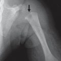
Fig. 5.34a–c Transient dislocation of the patella. (a) Lateral subluxation of the patella with an avulsion fraction off the medial pole. Bone contusions in the medial pole of the patella (arrow in b) and on the lateral condyle of the distal femur (arrow in c) from impaction by the patella.Fig. 5.35 Radial head dislocation. A line drawn through the center of the long axis of the proximal radius does not bisect the center of the capitellum.Fig. 5.36a–d Legg-Calvé-Perthes disease. Avascular necrosis (AVN) at the proximal epiphysis of the left femur in a patient with Legg-CalvéPerthes disease. Mild increased sclerosis on presentation (a) with decreased T1-weighted signal intensity (b) and increased T2-weighted signal intensity on MRI (c), as well as increased sclerosis 6 months later (d).Fig. 5.37a, b Juvenile idiopathic arthritis and sacroiliitis. Bone marrow edema on T2-weighted imaging (arrow in a) at the right sacroiliac joint with enhancement on T1-weighted imaging (b).
Table 5.26 Joints: congenital radial head dislocation
Diagnosis
Comments
Noonan syndrome
Male Turner syndrome. Multisystem involvement. Anterior bowing of the sternum, genu valgum, finger anomalies, scoliosis, vertebral anomalies, Klippel-Feil syndrome.
Short tubular bones particularly at the forearms, cone-shaped epiphyses of the phalanges and metacarpals, premature epiphyseal fusion of hands and feet, large great toes.
Auriculoosteodysplasia
Dysplasia of the radiocapitellar joint, ± radial head dislocation. Characteristic ear shape and short stature.
Hypoplastic clavicle, scoliosis, joint subluxations, long fingers and toes. Dense skull base, under-pneumatization of skull, short thumbs and great toes.