Abstract
Klippel-Trénaunay-Weber syndrome (KTWS) is characterized by a triad of cutaneous hemangiomas, hemihypertrophy, and vascular abnormalities. The prevalence of KTWS is 1 : 100,000. The exact pathophysiology and genetic etiology of the disorder are unknown. Prenatal diagnosis using ultrasound has been reported. Diagnosis is based on limb hypertrophy with the association of subcutaneous cystic lesions. Postnatal treatment consists of symptomatic management.
Key Words
Klippel-Trénaunay-Weber syndrome, hemihypertrophy, vascular abnormalities, subcutaneous cystic lesions
Introduction
Klippel-Trénaunay-Weber syndrome (KTWS) is characterized by a triad of cutaneous hemangiomas, hemihypertrophy, and vascular abnormalities. This triad of anomalies was first described by Klippel and Trénaunay in 1900. Parkes-Weber described an additional case 18 years later that had the triad of findings described by Klippel and Trénaunay and an arteriovenous malformation. The exact pathophysiology and genetic etiology of the disorder are unknown. Treatment consists of symptomatic management.
Disease
Definition
The diagnosis of KTWS is based on clinical findings. The major clinical findings are cutaneous hemangiomas, vascular abnormalities, and hemihypertrophy. To assign a diagnosis of KTWS, two of the three major findings must be present.
Prevalence and Epidemiology
The prevalence of KTWS is 1 : 100,000. It occurs equally in males and females and has no racial predilection.
Etiology and Pathophysiology
The pathophysiology and genetic etiology are unknown. Although a few familial cases and individuals with de novo balanced translocations have been reported, KTWS is generally accepted to be a sporadic disorder with no clear hereditary pattern.
Manifestations of Disease
Clinical Presentation
Cutaneous Hemangioma.
Hemangiomas are usually of the nevus flammeus type but can also occur as cavernous hemangiomas or lymphangiomas. Hemangioma depth is variable and can be limited to the skin or extend deeper into the visceral organs. If significantly large, hemangiomas can lead to platelet sequestering and possible Kasabach-Merritt syndrome (thrombocytopenia and microangiopathic hemolytic anemia in small children with hemangiomas).
Vascular Abnormalities.
The most common vascular abnormality seen is varicose veins. Varicose veins may manifest as superficial or deep and can remain stable or expand in size. Both lymphedema and pain have been associated with the varicosities. Arteriovenous malformations are less common vascular abnormalities in this syndrome.
Hemihypertrophy.
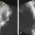
Hemihypertrophy usually occurs in extremities. Limb hypertrophy can manifest as bony or soft tissue involvement ( Figs. 131.1–131.3 ).


Occasional Findings.
Occasional findings include developmental delay, lymphatic obstruction, neural tube defect, hypospadias, polydactyly, syndactyly, oligodactyly, hyperhidrosis, hypotrichosis, ulceration, thrombophlebitis, angiosarcoma, pulmonary embolism, congestive cardiac failure, gangrene of the affected limb, and cellulitis.
Imaging Technique and Findings
Ultrasound.
Prenatal ultrasound (US) diagnosis of KTWS has been reported at 15 weeks’ gestation. Diagnosis is based on limb hypertrophy with the association of subcutaneous cystic lesions (see Fig. 131.1 ). Doppler interrogation of these subcutaneous cystic lesions failed to exhibit internal arterial or venous blood flow suggesting that either the vascular flow rates were too low for detection or lymphangiomas constituted most of the cystic mass. Other features noted on prenatal US consist of cardiac failure, polyhydramnios, Kasabach-Merritt syndrome, and fetal hydrops. Three-dimensional US has been used for additional assessment of leg width difference (see Fig. 131.3 ).
Magnetic Resonance Imaging.
Fetal magnetic resonance imaging (MRI) has been reported to be a useful adjunct testing modality in suspected cases. MRI aids in the characterization of soft tissue lesions and vascular abnormalities seen in KTWS.


