• The immediate subglottic trachea has a normal diameter but expands as it passes to the carina (and continuing into the major bronchi) • It is often associated with tracheal diverticulosis, recurrent lower respiratory tract infections and bronchiectasis
Large airway disease
TRACHEAL DISORDERS
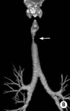
POST-TRAUMATIC STRICTURES
TRACHEAL DISORDERS
AMYLOIDOSIS
TRACHEOBRONCHOMEGALY (MOUNIER–KUHN DISEASE)
Definition
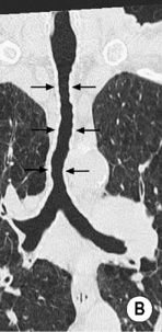
 atrophic mucosa prolapses between the cartilage rings giving the trachea a corrugated outline (this may become exaggerated to form sacculations)
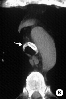
 it typically involves the trachea above the level of the thoracic inlet
 it involves 1.5–2.5cm of tracheal wall
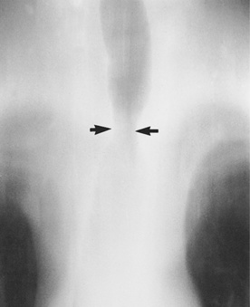
 it is also seen with TB, rhinoscleroma and necrotizing invasive aspergillosis
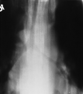
 it is characterized by gas within a cavitated hilar or mediastinal lymphadenopathy
 infection and trauma are other causes
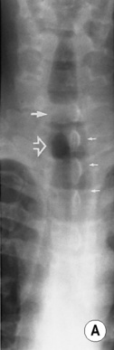
 a smoothly marginated intraluminal polyp (hamartomas and lipomas may demonstrate fat attenuation)
 it is often sessile and eccentric resulting in asymmetrical luminal narrowing
  can be polypoid and mostly intraluminal (with mediastinal extension seen in 30–40%)
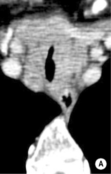
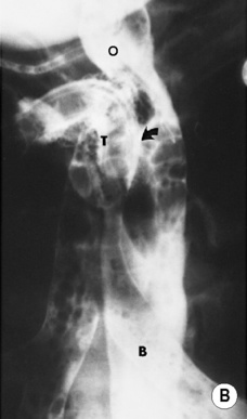
 it usually involves the larynx – occasionally extension into the trachea and proximal bronchi is seen
 although benign it may undergo transformation to a squamous cell carcinoma
 It is seen in association with tracheobronchomegaly, COPD, relapsing polychondritis and following trauma
 a coronal tracheal diameter significantly larger than the sagittal diameter (producing a lunate configuration)
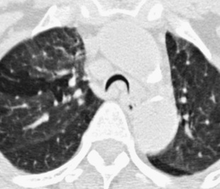
 nodular or polypoid lesions may be seen on the inner airway contour
  luminal stenosis may affect any main, lobar or segmental bronchus
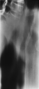
 usually there is a symmetrical subglottic stenosis – with disease progression the distal trachea and bronchi may become involved
 early sparing of the posterior tracheal wall (circumferential involvement with advanced disease)
  the trachea may become flaccid with considerable collapse at expiration
  fibrotic cartilaginous ring destruction may cause stenosis
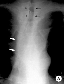
 dystrophic calcification or ossification is frequently present
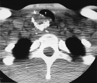
 the narrowing usually affects the whole intrathoracic trachea (with an abrupt return to a normal calibre at the thoracic inlet)
  frequently there is calcification of the tracheal cartilage rings
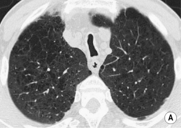
 diameters > 2.4 and 2.3cm for the right and left main bronchi, respectively
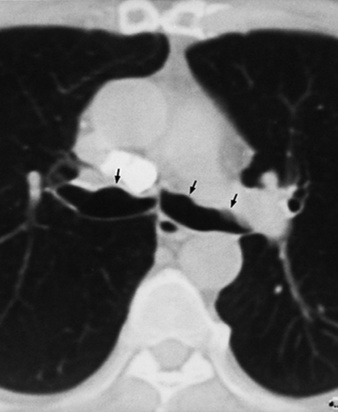
 the nodules contain heterotopic bone, cartilage and calcified acellular protein matrix with normal overlying bronchial mucosa
  M>F (usually > 50 years old)
 nodules may protrude from the anterior and lateral luminal walls into the lumen (usually with foci of calcification)
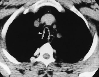
 the mechanisms include:

 sputum
  haemoptysis
  digital clubbing
 overinflation is often present with generalized disease (atelectasis can be seen with localized forms)
  thickened bronchial walls:


