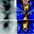
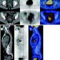
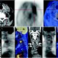
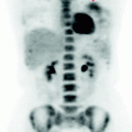
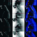
Fig. 68.1
At the CT there is evident massive opacification of the left hemithorax with marked attraction of mediastinal structures and pleural effusion
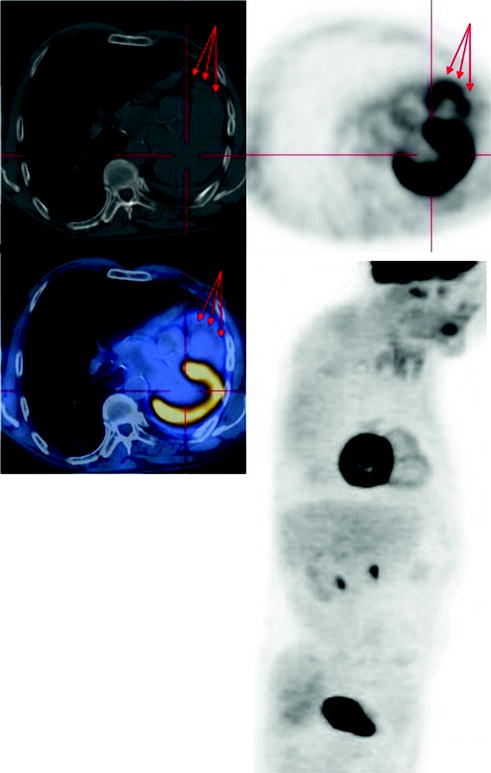
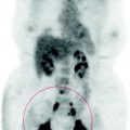
Fig. 68.2


The PET and axial MIP reconstruction image showed right ventricular hypertrophy and secondary pulmonary hypertension to the atria. No injuries to report a recurrence
Stay updated, free articles. Join our Telegram channel

Full access? Get Clinical Tree


