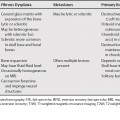
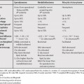
146 Classically, lesions involving the spinal canal have been approached by location. The division into extradural (outside the dural sac), intradural-extramedullary (within the dural sac, but outside the spinal cord) and intramedullary (within the spinal cord or filum terminale) has been the classic radiologic grouping. Extradural neoplasia is most common, and are often due to metastases involving the vertebrae, being followed by intradural-extramedullary tumors, with nerve sheath tumors and meningiomas being the most common, and lastly by the intramedullary tumors, which are predominantly astrocytomas and ependymomas. Degenerative etologies, including disc bulges, herniations, migratory fragments, facet arthorpathy, and ligamentum flavum redundancy, are the most common epidural lesions, exerting extrinsic compression on the thecal sac and nerve roots (see Chapter 149
Lesions within the Spinal Canal
Epidural Lesions
![]()
Stay updated, free articles. Join our Telegram channel


Full access? Get Clinical Tree


