and Lori Ann Lewis1
(1)
Clinical Research Center of North Texas, Denton, TX, USA
Abstract
Even though a technologist may follow positioning guidelines perfectly, the resulting study may be far from ideal because of artifacts or structural changes in the patient’s anatomy. Of the various skeletal regions of interest that may be studied with DXA, the lumbar spine is the region most commonly affected by artifact or structural change. The technologist obviously cannot correct structural changes in the patient’s skeleton, but it is imperative that they be recognized. Some structural changes or alterations will affect the measured BMD. Others may provide a possible explanation for the BMD value. Some artifacts are immediately recognizable while others are not. Any external artifacts should be removed if possible. If the artifact cannot be removed, the artifact should be identified and any possible effect on the measured BMD noted. Some explanations for structural changes or artifacts may be found in the patient’s history. The identification of others however is based purely on having seen it before. To that end, the DXA studies that follow reveal a variety of artifacts and structural changes encountered in clinical practice.
Even though a technologist may follow positioning guidelines perfectly, the resulting study may be far from ideal because of artifacts or structural changes in the patient’s anatomy. Of the various skeletal regions of interest that may be studied with DXA, the lumbar spine is the region most commonly affected by artifact or structural change. The technologist obviously cannot correct structural changes in the patient’s skeleton, but it is imperative that they be recognized. Some structural changes or alterations will affect the measured BMD. Others may provide a possible explanation for the BMD value. Some artifacts are immediately recognizable while others are not. Any external artifacts should be removed if possible. If the artifact cannot be removed, the artifact should be identified and any possible effect on the measured BMD noted. Some explanations for structural changes or artifacts may be found in the patient’s history. The identification of others however is based purely on having seen it before. To that end, the DXA studies that follow reveal a variety of artifacts and structural changes encountered in clinical practice.
Fig. 11-1.
This is a 67-year-old Caucasian woman. Height 64.2 in., weight 150.3 lb. The PA lumbar spine bone density study is shown in Fig. 11.1A. There is disc space collapse with marked endplate sclerosis at L3–L4 which affects the BMD at those levels and by extension, the L1–L4 BMD. Note that the BMDs at L1 and L2 are much lower than those at L3 and L4 because of the changes at L3 and L4. While the BMDs at L1 and L2 result in an L1–L2 BMD that would be considered osteoporotic, the L1–L4 BMD would be interpreted as normal if the structural changes and their effect on the L1–L4 BMD are not recognized. This patient’s left proximal femur study is shown in Fig. 11.1B. The bone density at the femoral neck is clearly osteoporotic and in keeping with the L1–L2 BMD in the spine. These studies were performed on a GE Lunar Prodigy.
Fig. 11-2.
This is a 70-year-old Caucasian woman. Height 63.6 in., weight 147.5 lb. There are pedicle screws clearly seen at L4 and L5. Although this is very dense material, the BMD at L4 is not markedly elevated compared to L3 because the analysis software of this DXA device identified these densities as nonphysiologic. They are, therefore, excluded from consideration in the analysis. However, the BMD at L4 cannot be considered valid. Note that the BMD at L1 is markedly lower than L2 or L3. In the author’s experience, this can be seen about 20 % of the time in the absence of any pathology. If the L1–L3 BMD was used, however, the low value at L1 would affect the L1–L3 BMD. Because this leaves only 2 vertebrae out of the 4 on which to base a diagnosis, greater weight should be given to any findings in the proximal femur. This study was performed on a GE Lunar Prodigy.
Fig. 11-3.
This is a 74-year-old Caucasian woman. Height 67.2 in., weight 128.2 lb. The PA lumbar spine study is shown in Fig. 11.3A. There is marked rotoscoliosis seen along with disc space collapse at L3–4 and L4–5. The sclerotic changes at L3–L4 and L4–L5 will increase the L1–L4 BMD and the corresponding T-score. If the L1–L4 T-score was used for diagnosis, the patient would be considered normal. The L1–L2 T-score is osteopenic. Recall, however, that rotation of the vertebrae tends to decrease the measured BMD. There is clearly rotation here. This patient’s dual femur study, shown in Fig. 11.3B, reveals a mean total hip T-score of −2.7, compatible with a diagnosis of osteoporosis. These studies were performed on a GE Lunar Prodigy.
Fig. 11-4.
This is a 62-year-old Caucasian woman. Height 63 in., weight 123.8 lbs. In the PA lumbar spine study shown in Fig. 11.4A, there appears to be disc space narrowing and a marked sclerotic change in the end plates at L1 and L2. The BMD at L1 is markedly increased as a result. It is reasonable to assume that there is some effect at L2 as well, leaving only L3 and L4 suitable for analysis. On this patient’s VFA image seen in Fig. 11.4B, osteophytic spurring, end plate sclerosis, and disc space narrowing are clearly seen at L1–L2. These studies were performed on a GE Lunar Prodigy.
Fig. 11-5.
This is a 36-year-old Caucasian woman. Height 61.7 in., weight 118 lbs. She underwent a surgical menopause at age 27 and only very briefly utilized hormone therapy. The PA spine bone density image seen in Fig. 11.5A shows nonphysiologic densities at L4 and L5, leaving L1–L3 suitable for analysis. The VFA image in Fig. 11.5B clearly shows the pedicle screws in the lower lumbar spine. These studies were performed on a GE Lunar Prodigy.
Fig. 11-6.
This is a 77-year-old Caucasian woman. Height 64.9 in., weight 168.2 lb. The PA lumbar spine image in Fig. 11.6A shows severe rotoscoliosis with the expected facet sclerosis. None of the vertebrae are suitable for analysis. Instead, the proximal femur study shown in Fig. 11.6B should be used. These studies were performed on a GE Lunar Prodigy.
Fig. 11-7.
This is a 69-year-old Caucasian man. Height 68.1 in., weight 164.6 lbs. Notice the unusual appearance of the lower lumbar spine in the PA spine bone density, particularly at L5. The posterior elements are not seen, and there appears to be a mass to the right of the spine. The patient’s history revealed that he had undergone a laminectomy and fusion at L5–S1. Thus, the posterior elements were missing because they had been surgically removed and the suspected mass was actually the fusion mass. These studies were performed on a GE Lunar Prodigy.
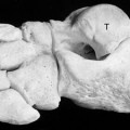
Fig. 11-8.


Related posts:
Stay updated, free articles. Join our Telegram channel
Full access? Get Clinical Tree
