Limited Vascular Compression Ultrasound for Deep Venous Thrombosis (DVT)


KEY IMAGES


Listen


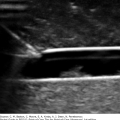
Start just below the inguinal ligament. Locate the common femoral vein (CFV) and femoral artery (FA). The great saphenous vein (GSV) enters from the medial side.


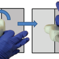
Hand position and anatomy


Figure 10-1


Ultrasound


Ultrasound with compression


Split of the common femoral vein into the deep femoral vein (DFV) and superficial femoral vein (SFV), and common femoral artery (CFA) into deep femoral artery (DFA) and superficial femoral artery (SFA).


Hand Position and Anatomy


Figure 10-2


Ultrasound


Figure 10-3


Ultrasound with Compression


Figure 10-4


Popliteal vein (PV) superficial to the popliteal artery (PA), with visible femoral condyle (FC)


Hand Position and Anatomy


Figure 10-5


Ultrasound


Ultrasound with Compression


Popliteal vein trifurcation into the peroneal, anterior, and posterior tibial veins


Hand Position and Anatomy

Only gold members can continue reading. Log In or Register to continue

Stay updated, free articles. Join our Telegram channel

Jul 6, 2019 | Posted by in ULTRASONOGRAPHY | Comments Off on Limited Vascular Compression Ultrasound for Deep Venous Thrombosis (DVT)

Full access? Get Clinical Tree
Get Clinical Tree app for offline access