Presentation and Presenting Images
( ▶ Fig. 76.1, ▶ Fig. 76.2, ▶ Fig. 76.3)
A 70-year-old female with a history of left mastectomy for papillary carcinoma 4 years ago, presents for routine screening mammography of her right breast.
76.2 Key Images
76.2.1 Breast Tissue Density
There are scattered areas of fibroglandular density
76.2.2 Imaging Findings
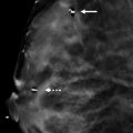
A linear density (oval) is seen on the mediolateral (MLO) view ( ▶ Fig. 76.4) and the lateromedial (LM) view ( ▶ Fig. 76.5), but not on the craniocaudal (CC) view. The CC view is normal. This density was not seen on prior mammograms (not shown).
76.3 BI-RADS Classification and Action
Category 0: Mammography: Incomplete. Need additional imaging evaluation and/or prior mammograms for comparison.
76.4 Diagnostic Images
( ▶ Fig. 76.6, ▶ Fig. 76.7, ▶ Fig. 76.8)
76.4.1 Imaging Findings
A linear density (oval) is seen on the MLO spot-compression view ( ▶ Fig. 76.7). The patient had a repeat MLO view ( ▶ Fig. 76.8) and an LM digital breast tomosynthesis (DBT) view. The additional imaging showed no suspicious findings. The patient may return to annual mammography. The appropriate management would have been to repeat the MLO view rather than performing a MLO spot-compression view.
76.5 BI-RADS Classification and Action
Category 1: Negative
76.6 Differential Diagnosis
Pseudolesion, or summation artifact, due to undercompression: In this case, the finding is noted in the MLO and LM views but not in the CC view. The length of the finding is less on the LM view than the MLO view, consistent with a pseudolesion due to undercompression. The finding is not reproduced on the DBT movie.
Postreduction scar: The finding is linear; however, the location is too high for a postreduction scar. The finding is not reproduced on the DBT movie.
Foreign body: The finding is not well seen on all views and is not reproduced on the tomosynthesis movie.
76.7 Essential Facts
Although, breast cancer may present as a one-view mammographic finding, frequently one-view findings are due to overlapping breast tissue secondary to poor positioning and undercompression.
One-view findings are commonly a summation artifact.
With compression of the three-dimensional (3D) breast on a two-dimensional (2D) view, overlap is inevitable. Overlap occurs in breasts of all densities but is more common in dense breasts.
Sometimes when the standard two mammographic breast views are compared, the radiologists can ascertain that the asymmetry spreads out in the other view in what Brenner has termed the “schmear” sign.
76.8 Management and Digital Breast Tomosynthesis Principles
With DBT, the image data set is less vulnerable to summation artifacts than conventional mammography.
The use of both conventional mammography and DBT has been shown to reduce recall rates for women without breast cancer. DBT would have prevented additional imaging in this case.
DBT has been shown to be less likely than conventional mammography to require recall for asymmetries, focal asymmetries, and architectural distortions. This is due to DBT’s ability to eliminate overlap.
76.9 Further Reading
[1] Brenner RJ. Asymmetric densities of the breast: strategies for imaging evaluation. Semin Roentgenol. 2001; 36(3): 201‐216 PubMed
[2] Ciatto S, Houssami N, Bernardi D, et al. Integration of 3D digital mammography with tomosynthesis for population breast-cancer screening (STORM): a prospective comparison study. Lancet Oncol. 2013; 14(7): 583‐589 PubMed
[3] Giess CS, Frost EP, Birdwell RL. Interpreting one-view mammographic findings: minimizing callbacks while maximizing cancer detection. Radiographics. 2014; 34(4): 928‐940 PubMed

Fig. 76.1 Right craniocaudal (RCC) mammogram.
Stay updated, free articles. Join our Telegram channel

Full access? Get Clinical Tree


