Clinical Presentation
The patient is a 52-year-old female with symptoms of progressive decrease in strength in both legs and urinary incontinence. There is no history of antecedent trauma. No recent infection or vaccinations. She had a similar bout of leg weakness and urinary incontinence, although much less severe, 25 years ago when she was pregnant. These symptoms went away approximately 3 months postpartum. On physical examination, the patient has a decrease in sensation from approximately T2 inferiorly. Her gait is slightly broad-based and there is symmetric, mild lower extremity weakness.
Imaging Presentation
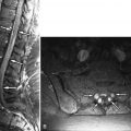
Sagittal T1- and fat-saturated, T2-weighted magnetic resonance (MR) images reveal an oblong intraspinal mass with increased T1 signal intensity and low signal intensity on the fat-saturated acquisition. A nonfat-saturated, T2-weighted sequence (not pictured) revealed the mass to be of increased signal intensity. The mass has the imaging characteristics of fat and represents an intradural lipoma ( Fig. 43-1 ) .

Discussion
Intradural lipomas of the spinal canal are rare and comprise less than 1% of all primary tumors of the spine. Vertebral, dermal, and renal abnormalities are not a feature of intradural lipomas because they, along with other fatty neoplasms, are associated with spinal dysraphism. Of all fatty neoplasms of the spinal canal, intradural lipomas account for 4% of the tumors, lipomyelomeningoceles for 84%, and lipomas of the filum terminale for 12%. Intradural spinal lipomas have nearly equal gender preference with a very slight female predominance. They most commonly manifest in the second and third decades of life (55%), and to a lesser extent in the first year of life (24%) and fifth decade of life (16%). The thoracic region is most commonly involved in adults (32%), followed by the cervicothoracic region (24%), and cervical region (13%). Intradural spinal lipomas are usually posterior (67%) or posterolateral (23%) to the spinal cord. The cervical region is most commonly involved in children.
There is debate as to the embryology of intradural spinal lipomas. It is thought that lipomas develop when there is premature dysjunction of the cutaneous ectoderm from the forming neural tube. The surrounding mesenchyme migrates underneath the ectoderm into the neural tube and adheres to the primitive ependyma, which induces it to dedifferentiate into fat. Normally, the mesenchyme migrates into the space between the neural tube and superficial ectoderm and differentiates into the vasculature for the spinal cord, meninges, and vertebral column. Histologically, intradural spinal lipomas consist of mature fat cells separated by connective tissue strands and are partially to fully encapsulated. The lipoma is admixed with nerve bundles that are often located at the periphery, which suggests secondary entrapment of adjacent nerve roots. Although these lipomas are not considered neoplastic, they can demonstrate autonomous, slow growth. The fat of these lipomas is metabolically active like fat in the rest of the body. Growth of these lipomas can be seen with an increase in body fat and are associated with the metabolic changes of pregnancy.
The clinical course of patients with intradural spinal lipomas is one of slow progression of myelopathic symptoms. Fifty-five to sixty-eight percent of patients become symptomatic in the first three decades of life. Patients often present with a long history of disabilities that rapidly progress prior to them seeking medical attention. Symptoms include spinal pain, sensory changes, loss of positional sense, gait disturbance, weakness, hypotonia, and incontinence. Early gait disturbance is caused by dorsal column dysfunction (secondary to the posterior location of most lipomas) rather than weakness in the legs. The most frequently reported symptoms are numbness or spastic weakness in the extremities. Radicular pain is uncommon.
Imaging Features
Magnetic resonance imaging (MRI) is the imaging modality of choice. The mass is readily identified, as are its location and effect on the spinal cord, other nervous tissue, and vasculature. Intradural lipomas often appear as round or lobulated masses that display signal characteristics of normal fat. The spinal canal may demonstrate widening in the region of the mass because of chronic pressure erosion. The mass is of increased signal intensity on T1- and T2-weighted acquisitions, and displays low signal intensity on both sequences when a fat-saturation technique or short tau inversion recovery (STIR) technique is applied (see Fig. 43-1 ). Lipomas do not demonstrate contrast enhancement. If the fatty mass demonstrates a region of contrast enhancement, then a more ominous etiology such as liposarcoma should be entertained. The adjacent spinal cord may be compressed or appear stretched. There may be increased T2 signal in the intrinsic cord compatible with cord ischemic change or myelomalacia. Computerized tomography (CT) demonstrates spinal lipomas as a lobulated/ovoid hypodense (fat density Hounsfield unit) mass ( Fig. 43-2 ) . CT does not identify the relationship of the mass to the spinal cord and nerve roots as well as MRI. Myelography does not identify the mass but demonstrates the effect of the mass on the dural sac and spinal cord. When viewed in a plane parallel to the mass and spinal cord, an intradural extramedullary mass will appear as a negative defect in the spinal column with widening of the contrast-filled space immediately cephalad and caudad to the mass, between the dural sac and the negative defect from the spinal cord ( Fig. 43-3 ) . The addition of CT to myelography gives only marginally more benefit than CT alone. Ultrasound can be used in infants suspected of having a spinal lipoma. Ultrasound, unlike CT, does not use ionizing radiation, and it is unnecessary to sedate an infant for an ultrasound examination, unlike MRI. A lipoma will appear as an echogenic mass.


