KEY FACTS
Imaging
- •
Portal vein stenosis
- ○
Elevated portal vein velocity > 125 cm/s at anastomosis
- –
Portal vein velocity normally < 60 cm/s
- –
- ○
Anastomotic:preanastomotic velocity ratio
- –
Stenosis: > 3.0
- –
Normal: ~ 1.5
- –
- ○
- •
Portal vein thrombosis
- ○
No detectable flow in portal vein
- ○
May see echogenic thrombus in portal vein, or portal vein may appear markedly hypoechoic or even anechoic
- ○
May see enlarged hepatic arteries
- –
Hepatic arteries compensate for portal vein thrombosis due to hepatic arterial buffer response
- –
- ○
Bidirectional low-amplitude flow in portal vein may precede portal vein thrombosis
- ○
Pathology
- •
Portal vein stenosis
- ○
Occurs at site of anastomosis
- ○
- •
Portal vein thrombosis
- ○
Associated with surgical technique
- ○
Hypercoagulable state
- ○
Clinical Issues
- •
Portal vein stenosis typically occurs at anastomosis
- •
Depending on degree of stenosis, may require balloon angioplasty
- •
Portal vein stenosis and thrombosis are relatively rare, occurring in 1-2.7% of liver transplants
Scanning Tips
- •
In chronic or longstanding portal vein stenosis, may see portal vein aneurysm downstream from anastomosis and helical flow in dilated portion of portal vein
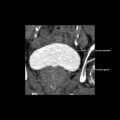
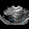
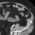
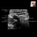
 of the portal vein at the level of the anastomosis in a patient with a liver transplant.
Related posts:


Stay updated, free articles. Join our Telegram channel

Full access? Get Clinical Tree


