Chapter 16
Liver trauma
Ersan Altun1, Mohamed El-Azzazi1,2,3,4, and Richard C. Semelka1
1The University of North Carolina at Chapel Hill, Department of Radiology, Chapel Hill, NC, USA
2University of Dammam, Department of Radiology, Dammam, Saudi Arabia
3King Fahd Hospital of the University, Department of Radiology, Khobar, Saudi Arabia
4University of Al Azhar, Department of Radiology, Cairo, Egypt
- Blunt and penetrating trauma.
- Blunt trauma is more common.
- Blunt trauma most commonly involves the right hepatic lobe as it constitutes the greatest volume of the liver. The posterior segment is the most commonly involved segment as it is susceptible to blunt impact (occasionally penetrating) from the ribs and spine and relative fixation of the liver by the coronary ligaments.
- Blunt trauma is more common.
- CT and MRI findings of hepatic injury:
- Laceration or fracture through the hepatic parenchyma:
- Contrast enhanced examinations are essential for the evaluation both on CT and MRI.
- Appear as irregular linear, branching, or rounded areas of low attenuation within the normally enhancing liver on contrast enhanced CT images. These areas may not be detected on unenhanced CT images. They demonstrate mild to moderately high signal on precontrast T2-WI and low signal on T1-WI on MRI.
- High-attenuation foci of freshly clotted blood may be seen in the areas of laceration on unenhanced and contrast enhanced CT images. These areas demonstrate variable MRI signal according to the age of blood products; however, they usually demonstrate high signal on precontrast T1-weighted images.
- Lacerations commonly parallel the hepatic or portal venous vasculature and often extend to the periphery of the liver. They may have a configuration that has been termed the bear claw pattern due to its radiating, parallel, and jagged appearance.
- On occasion, hepatic lacerations may demonstrate a branching pattern that superficially simulates the appearance of dilated bile ducts.
- Lacerations extending into the perihilar region of the liver have an increased incidence of bile duct injury.
- Contrast enhanced examinations are essential for the evaluation both on CT and MRI.
- Intraparenchymal hematoma:
- They tend to be rounded or oval in configuration, and often display a central high-attenuation area of clotted blood surrounded by a larger low-attenuation region of lysed clot and contused liver parenchyma on unenhanced and contrast enhanced CT.
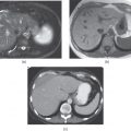
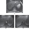
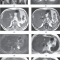
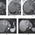
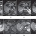
- Hematomas demonstrate variable MRI signal on precontrast T1- and T2-WI according to the age of blood products. They commonly show heterogeneously increased T2 signal containing low signal intensity areas and heterogeneously low T1 signal containing high signal intensity areas (Figures 16.1 and 16.2). Layering blood products may be seen.
- They tend to be rounded or oval in configuration, and often display a central high-attenuation area of clotted blood surrounded by a larger low-attenuation region of lysed clot and contused liver parenchyma on unenhanced and contrast enhanced CT.
- Subcapsular hematoma:

Stay updated, free articles. Join our Telegram channel
- Laceration or fracture through the hepatic parenchyma:

Full access? Get Clinical Tree


