Local Tumor Recurrence
KEY FACTS
Terminology
Imaging
IMAGING
General Features
Ultrasonographic Findings
 Presence of discrete soft tissue mass at surgical site is best diagnostic clue
 Variable echogenicity and vascularity of recurrent tumor but mirrors that of primary tumor
 Associated abnormality
 US useful at detecting bone tumor recurrence
 Most bone tumor recurrences occur in soft tissue and not in bone
 Most recurrences lie close to prosthetic or allograft implant
 Discrete soft tissue mass is best diagnostic clue
Imaging Recommendations
 US, as it is easily available, does not require contrast injection and is not subject to metallic artifact
 For high-grade bone or soft tissue sarcoma (STS), follow-up at 3 months after surgery, then every 6 months for 2 years, then yearly thereafter for 5 years
 For low-grade bone or STS, follow-up at 6 months after surgery, then yearly thereafter for 3 years
 However, still not clear whether routine surveillance actually improves survival
 Same protocol is used for both bone and soft tissue recurrence
 Examine entire section of limb from which primary tumor was resected (e.g., thigh, leg, arm, or forearm)
 Use parallel series of longitudinal sweeps (with transducer aligned transversely) from proximal to distal along affected limb segment
 Examine patient in both supine and prone positions to ensure that entire circumference of operated segment of limb is examined
 Look for any discrete soft tissue mass, particularly along scar
 Consider likelihood of postoperative, nontumoral masses
![]()
Stay updated, free articles. Join our Telegram channel

Full access? Get Clinical Tree


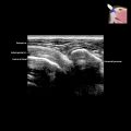
 . A nodule
  at the posterior aspect of the flap is slowly increasing in size.
 . Features are highly suspicious for local recurrence.
 suggestive of tumor recurrence.
 at the anteromedial aspect of prosthetic bone junction
  with irregular underlying osteolysis
  . Percutaneous biopsy confirmed local recurrence. MR and CT would have been limited by metallic artifact while US clearly shows recurrence.