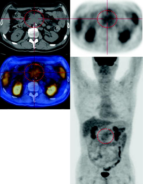
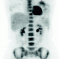
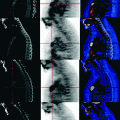
Fig. 20.1
In the celiac-mesenteric intercavo-aortic region, CT-PET demonstrates a solid mass with parenchymal density, uneven with irregular margins, not separable from the surrounding structures and characterized by limited concentration of FDG
Absence of significant focal areas of abnormal metabolism in the remaining body segments examined.
20.4 Conclusions
The PET scan suggests recurrence of mucinous adenocarcinoma of the colon with limited metabolism of glucose. (See Fig. 20.2).
