Abstract
The lung is an organ where integration of data from FDG PET, the CT, and the clinical history is particularly important. There are many FDG-avid lung lesions which will be determined to be benign or malignant only after correlation with CT findings and the clinical history. FDG PET/CT plays an important role in staging of lung cancer and determining if surgical resection is possible.
Keywords
FDG, PET/CT, lung, solitary pulmonary nodule, lung cancer
The lung is an organ where the integration of findings on 18F-fluorodeoxyglucose positron emission tomography (FDG PET), findings on computed tomography (CT), and the clinical scenario is particularly important to arrive at the best conclusions. Many FDG-avid lung lesions will be determined to be malignant or benign only after correlation with CT findings and the clinical history. Non-FDG-avid lung lesions may also be malignant and need to be recognized on the CT component of the study.
It is useful to consider multiple differential diagnoses for pulmonary lesions. The lung is a common site of malignancy, including metastases, primary lung cancers, neuroendocrine tumors/carcinoid, and lymphoma. There are many benign etiologies of lung lesions: infectious (such as granulomas, pneumonia, abscesses), vascular (embolic, aterio-vascular malformations), autoimmune (rheumatoid nodules, Wegener/granulomatosis with polyangiitis), benign neoplasms (hamartomas), congenital (bronchogenic cysts), and traumatic (pneumatocele) lesions.
Solitary Pulmonary Nodules
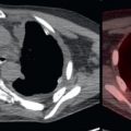
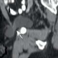
A solitary pulmonary nodule (SPN) is a round intraparenchymal lung lesion less than 3 cm in greatest diameter, without adjacent atelectasis or local suspicious lymph nodes. I mention this definition, since many studies are ordered with a clinical indication of SPN, without actually being an SPN. FDG PET/CT has been estimated to have about 80% accuracy in defining an SPN greater than 8 mm as benign or malignant. This number will vary depending on where you live, as regions with endemic granulomatous diseases will have a greater prevalence of benign SPNs. This relatively high accuracy led to the evaluation of SPNs as one of the early approved applications for FDG PET ( Fig. 8.1 ). However, an FDG-avid SPN is not always malignant ( Fig. 8.2 ), and some primary lung malignancies such as adenocarcinoma in situ, minimally invasive adenocarcinoma ( Fig. 8.3 ), well-differentiated adenocarcinoma ( Fig. 8.4 ), and low-grade carcinoids may not be FDG avid. All SPNs should be evaluated by tissue sampling or follow-up imaging. FDG PET/CT may help distinguish between these two choices, as a non-FDG-avid SPN may favor follow-up imaging, while FDG-avid SPN favors immediate tissue sampling.


Multiple Pulmonary Nodules
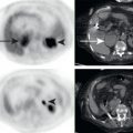
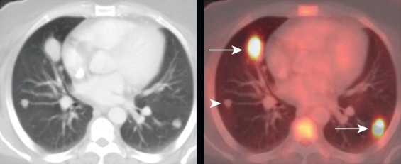
Multiple pulmonary nodules also have a wide differential diagnosis, including malignant (metastases, primary lung cancers, lymphoma) and benign (embolic, autoimmune, infectious) etiologies. Both the CT and FDG PET characteristics, as well as the clinical scenarios, should be considered when characterizing multiple pulmonary nodules. In a patient with a malignancy with propensity to metastasize to the lung, multiple round pulmonary nodules should be considered suspicious for lung metastases, whether FDG avid ( Fig. 8.5 ) or not ( Fig. 8.6 ). Likewise, in a patient with lymphoma, multiple round nodules in the lungs should be considered lung lymphoma until proven otherwise ( Fig. 8.7 ). Metastases are most commonly round nodules; thus the presence of multiple FDG-avid patchy nodules may suggest a different diagnosis ( Fig. 8.8 ). As can be seen, both the CT and FDG PET characteristics of multiple pulmonary nodules are important for optimal diagnosis.


Stay updated, free articles. Join our Telegram channel

Full access? Get Clinical Tree


