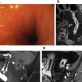
Colorectal cancer is the second most common cause of cancer-related death in Europe and the United States, and a major cause of mortality. Early detection of colorectal cancer and its precursors reduces mortality and morbidity, and a minimally invasive screening tool is essential for high patient acceptance and participation. To achieve this goal, computed tomographic colonography and magnetic resonance (MR) colonography have been introduced. A wide variety of methods of bowel preparation, colon distension, and imaging exists for MR colonography. This article presents an up-to-date overview of the status of MR colonography in screening for colorectal cancer, and its diagnosis.
Key points
- •
Magnetic resonance (MR) colonography is a potential technique for colorectal cancer screening because of its lack of ionizing radiation and high soft-tissue contrast, thus rendering exceptional extracolonic assessment.
- •
Prerequisites of MR colonography are a bowel preparation with fecal tagging and adequate colonic distension.
- •
The results of diagnostic performance of MR colonography are promising but heterogeneous, which hampers the use of this modality as a screening tool for colorectal cancer at present.
Introduction
Colorectal cancer is the second most common cause of cancer-related death in Europe and the United States, and is a major cause of mortality. In 2012, an estimated 143,460 new cases were diagnosed with colorectal cancer in the United States. The 5-year relative survival rate is 90.1%, if colorectal cancers are detected at a localized stage. However, when it has spread to regional lymph nodes and organs, the 5-year survival decreases to 69.2% and when it has metastasized to other than regional areas, survival decreases even further to 11.7%. Early detection of colorectal cancer is therefore essential for high survival rates. Moreover, colorectal cancer can be prevented, as precancerous adenomatous polyps can be detected and removed. It is estimated that screening for colorectal cancer can reduce colorectal mortality by more than 50%.
Different screening tools such as fecal occult blood tests (FOBT), barium contrast enema, sigmoidoscopy, and colonoscopy have been evaluated. Among these procedures, optical colonoscopy is the most accurate means for examining the colon, with high sensitivity and specificity regarding the detection of colorectal cancer and adenomatous polyps, with polypectomy reducing mortality rates of colorectal cancer. Nevertheless, moderate patient acceptance has been demonstrated for screening programs in comparison with other screening methods (ie, FOBT), the participation rate of colonoscopy is demonstrably lower (FOBT 47%, colonoscopy 22%).
In the 1990s the first attempts were made to develop an imaging-based minimally invasive technique for the detection of colorectal cancer and its precursors. The available structural examinations (ie, optical colonoscopy and sigmoidoscopy), were subject to patient discomfort, which led to the development of both computed tomography (CT) colonography and MR colonography. CT colonography was described first, and initial attempts were challenging because of time-consuming image reconstructions, disk-storage capacities, and the development of patient-preparation methods to avoid false-positive and false-negative findings. Since then tremendous progress has been made in spatial resolution and the examination and interpretation times of CT colonography, and it is nowadays part of daily clinical practice. Moreover, CT colonography has proved to be highly accurate in detecting colorectal cancer and large lesions of 10 mm and greater. In both average-risk and high-risk individuals, detection of precursors of colorectal carcinoma is high: per-patient sensitivity is 88% for advanced neoplasia of 10 mm or larger in screening populations and 96% for colorectal carcinoma in average-risk and high-risk populations, findings comparable with those of colonoscopy. This success has resulted in the recommendation of CT colonography as a screening tool for colorectal cancer by the American Cancer Society.
The major advantage over colonoscopy and sigmoidoscopy is that colonography (also known as virtual colonoscopy) lacks the need of sedation and a cathartic bowel preparation. Moreover, the risk of procedural complications is lower.
Substantially more data are available on the diagnostic performance of CT colonography in comparison with MR colonography. CT colonography has the advantage of shorter examination times, clinical availability, and lower costs. However, the major motivation to use MR colonography is the lack of ionizing radiation. This issue seems to be of lesser importance as the daily dose of radiation with CT colonography has been significantly reduced. Nonetheless, evaluation of the complete colon without the use of ionizing radiation would be most favorable, as ionizing radiation might be a concern when used for screening purposes. This aspect is especially important because screening by colonography most likely will not be limited to a single examination. Moreover, the excellent soft-tissue contrast of MR imaging has made MR colonography attractive for the evaluation of other abdominal abnormality (eg, inflammatory bowel disease, which is described in an article elsewhere in this issue by Jordi and colleagues).
The aim of this review is to describe the current status and potential for MR colonography in screening for colorectal cancer.
Stay updated, free articles. Join our Telegram channel

Full access? Get Clinical Tree


