The glenohumeral joint enables tremendous range of motion at the expense of stability. Functional stability is maintained by the synchronous coordination of complex static and dynamic structures. Symptomatic glenohumeral instability most often results from injury to the inferior labral-ligamentous complex, the primary passive stabilizer of the shoulder. This article reviews the structures important in glenohumeral stabilization and illustrates their normal appearances and the abnormalities associated with anterior, posterior, and multidirectional instability. These lesions are discussed in the context of therapeutic decision making.
The glenohumeral joint is one of the least anatomically constrained in the body, conferring an advantage in mobility but causing a propensity for instability. In patients with unstable shoulders, symptoms such as apprehension, looseness, slipping, or “going out” usually correlate with objective signs on physical examination. These clinical features may warrant imaging studies for lesion characterization, preoperative planning, and prognosis, and the detection of secondary degenerative change.
Historically, glenohumeral instability has been classified by both directionality (anterior, posterior, or multidirectional) and origin (traumatic or atraumatic). Trauma typically causes unidirectional instability and results from single inciting events (eg, fall on an out-stretched hand) or repeated high-load activities (eg, weight-lifting). After trauma, orthopedists can usually determine the predominant direction of instability based on history, symptoms, and signs, whereas atraumatic instability is more difficult to characterize. Causes include connective tissue disorders or repeated low-load activities (eg, swimming) that lead to generalized ligamentous laxity and multidirectional instability (MDI), often affecting both shoulders equally. Although trauma-related instability can be obvious clinically, shoulder hypermobility caused by atraumatic instability has proved difficult to distinguish from hypermobility caused by benign capsular laxity. The differentiation is important because true instability eventually leads to debilitation and progressive joint degeneration. Regardless of whether it is causing instability, capsular laxity remains a difficult imaging diagnosis.
This article first reviews the anatomic structures most important in glenohumeral stability and the most common instability patterns. Subsequently, the focus shifts to MR imaging findings in unstable shoulders.
Functional stability
The normal shoulder maintains stable alignment because of osseous congruity, labrocapsular integrity, and synergistic muscle balance. The osseous geometry of the glenohumeral joint contributes less to functional stability than in other joints, because the glenoid surface area is small and shallow compared with the humeral articular surface. Glenoid hypoplasia further reduces glenoid surface area and predisposes toward instability. For example, patients with an acquired or developmental deficiency of the posteroinferior glenoid rim have increased risk of posterior shoulder instability.
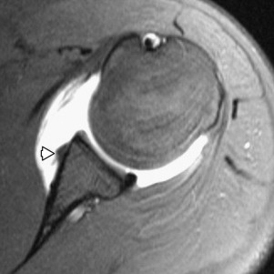
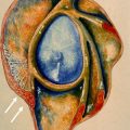
The glenoid labrum encircles the glenoid, enhancing both depth and surface area of the glenoid cavity, and consequently improving congruity of the two articular surfaces ( Fig. 1 ). The glenoid labrum is commonly described as a fibrocartilaginous structure; however, histologic examinations have shown that the labrum consists of parallel collagen fibers and dense fibrous connective tissue without chondrocytes, an appearance that corresponds to the morphologic structure of a tendon. The labrum functions as a “chock block” to resist translational forces during movement and, most importantly, acts as a point of attachment for the joint capsule, glenohumeral ligaments (GHLs), and biceps tendon. As a capsular attachment site, the labrum helps to create a suction seal. Negative intraarticular pressure boosts shoulder stability unless the seal is lost because of rotator cuff tear or labral tear. Compared with the anteroinferior labrocapsular complex, the anterosuperior labrocapsular complex is less important in shoulder stabilization. Superior labral lesions occur in patients with clinically stable shoulders and are often debrided without shoulder destabilization. Developmental variations of the anterosuperior labrum, due to a sublabral foramen or Buford complex ( Fig. 2 ), are not associated with shoulder instability.


The glenohumeral joint capsule has ligamentous thickenings ( Fig. 3 ) that are consistently shown on MR arthrography. The most important ligaments serve important biomechanical roles through limiting excessive translation and rotation of the humeral head. Numerous in vivo and in vitro studies have investigated the complex anatomic configurations and biomechanical contributions of these structures. The coracohumeral ligaments (CHLs), superior GHLs, and middle GHLs are less important than the inferior GHL. If the inferior GHL is attached to a labrum that is torn from the glenoid rim, it becomes incompetent and the shoulder becomes unstable.

On MR images, the CHL extends laterally from the dorsal base of the coracoid. The superior GHL courses parallel to the coracoid process from its labral attachment site and merges with the CHL in the rotator interval, reinforcing the biceps tendon sling (pulley). The CHL also branches posteriorly to envelope the supraspinatus and infraspinatus tendons, forming the semicircular humeral ligament (rotator cable). The middle GHL arises from the anterosuperior glenolabral rim with the superior GHL and courses caudally, merging with the subscapularis tendon near the lesser tuberosity ( Fig. 4 ). The labrum, middle GHL, and inferior GHL share strong developmental relationships. Based on cadaver studies, the middle GHL is absent in approximately 30% of patients. When present, it ranges in thickness from wafer-thin to cord-like. The cord-like middle GHL can be associated with absent anterosuperior labrum (Buford complex) (see Fig. 2 ). When the middle GHL is small or absent, there is often compensatory enlargement and superior takeoff of the inferior GHL. Although the CHL, superior GHL, and middle GHL make limited stabilizing contributions, many authors associate laxity of these structures with microinstability and MDI. Therefore, some surgeons tighten the rotator interval (capsulorrhaphy) in patients with MDI.

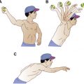
Compared with other capsuloligamentous structures, the inferior GHL contributes most to passive glenohumeral stabilization. The inferior GHL includes an anterior band (aIGHL) (see Fig. 4 ), posterior band, and intervening axillary pouch. The aIGHL resists anteroinferior translation of the humeral head and becomes taut when the shoulder is abducted and externally rotated (ABER position) ( Fig. 5 ). In most patients, the aIGHL arises from the labroglenoid rim inferior to the 3 o’clock position, though it may arise more superiorly. The aIGHL usually attaches to the labrum and glenoid rim, forming a sleeve of continuous tissue between the labrum, capsule, and periosteum. Lesions involving this periosteal sleeve are often associated with instability. The posterior band of the inferior GHL more commonly arises directly from the posteroinferior glenoid rather than the labrum. The humeral attachment of the inferior GHL complex is either (1) collar-like, in which the anterior and posterior bands of the inferior GHL fan out and create a ring-like insertion slightly caudal to the articular edge of the humeral head, or (2) V-shaped, in which the anterior and posterior bands of the inferior GHL come together and create a point-like insertion.

The dynamic stabilizers of the shoulder include the rotator cuff, deltoid, periscapular muscles and the biceps tendon (long head). During normal shoulder movements, these structures maintain a delicate contractile balance to center and compress the humeral head into the labroglenoid socket and counter the translational forces inherent in shoulder motion. This “compression concavity” mechanism is most important in the middle ranges of motion when the capsuloligamentous structures are lax.
Instability patterns: clinical perspective
Anterior Instability
The glenohumeral joint dislocates more commonly than any other, occurring in 11.2 per 100,000 persons per year. Most dislocations (90%) are directed anteriorly. The mechanism can involve falling on an outstretched arm or colliding with players during contact sports, such as American football or rugby. At the time of injury, the shoulder is often forced anteriorly in abduction, external rotation, and extension. A patient may not realize that the shoulder has dislocated and spontaneously relocated. First-time dislocations can be difficult to diagnose unless confirmed through reduction maneuvers or radiographs. In a recent study of young athletes, first-time trauma-related shoulder injuries were clinically categorized as subluxations in 85% of cases and as dislocations in only 15% of cases. Although structural damage is commonly associated with dislocations, the authors also reported a high rate of labral, capsular, and osseous lesions in their patients with anterior subluxations.
After traumatic shoulder dislocation (treated nonoperatively), the likelihood of repeat dislocation depends on patient age and the date of initial injury. Patients younger than 20 years have a higher risk of recurrent dislocation (>90% when subluxation events are included) than those older than 40 years (<10%). The likelihood of recurrent dislocation also depends on behavior modification. If an athlete returns to a collision sport, such as football and hockey, or a contact sport, such as lacrosse and basketball, the incidence of recurrent dislocation approaches 100% at the same level of competition. Older individuals are much more willing to modify behavior to avoid repeated dislocations, chronic anterior instability, and progressive functional disability.
Posterior Instability
Posterior instability is much less common than anterior instability. An incidence of 2% to 4% is generally accepted, but a higher incidence (11.6%) was reported more recently when recurrent posterior subluxation was taken into account. Traumatic posterior dislocation typically occurs after a blow to the upper arm when the humerus is flexed, adducted, and internally rotated (opposite to anterior dislocation). Bilateral posterior dislocation can occur after an epileptic seizure or high-voltage electric shock from the strong contraction of internal rotators.
MDI
MDI represents a complex condition without consistent diagnostic and treatment criteria. In 1980, Neer and Foster reported that patients treated surgically for MDI presented with predominantly inferior signs and symptoms. More recently, authors have recommended that the diagnosis of MDI requires symptoms and signs of instability in an inferior direction and at least one other direction (anterior or posterior). MDI often develops in the absence of trauma (atraumatic instability), involves both shoulders, and indicates joint hypermobility from generalized capsular laxity. Acquired causes may reflect repetitive overstretching activities (eg, swimming, gymnastics), proprioceptive imbalances, connective tissue disorders, or multifactorial conditions. Some authors argue that all shoulder instabilities show some degree of excessive multidirectional translation and, therefore, propose that the multidirectional descriptor should be dropped so that instabilities are described based only on the predominant direction of abnormal motion.
Imaging techniques
At the authors’ institution, referring physicians request the imaging modality. In patients with suspected shoulder instability, the cross-sectional modality could be CT, CT arthrography, MR imaging, or MR arthrography. It is extremely rare for a radiologist to change the request. Some orthopedists depend on nonarthrographic studies, but others strongly prefer arthrographic techniques unless recent acute trauma has occurred, in which case joint effusion, edema, and hemorrhage are usually present. MR arthrography is more commonly requested in younger individuals with clinical evidence of shoulder instability. Although certain referrers exclusively select MR, others are balanced in their choice of MR versus CT, depending on the suspicion of glenoid rim fracture and the likelihood of performing Latarjet reconstruction of the glenoid rim.
In the assessment of the labrocapsular complex, MR arthrography has shown sensitivities of 86% to 91% and specificities of 86% to 98%, whereas conventional MR imaging has shown sensitivities and specificities ranging from 44% to 100% and 66% to 95%, respectively. MR arthrography can add confidence in the diagnosis of labral tear compared with conventional MR imaging, even at 3T. A potential advantage of MR arthrography is ABER positioning (see Fig. 4 ). If the position can be tolerated, it may improve the detection of subtle anteroinferior labral tears, while also revealing tendon delamination involving the rotator cuff. Studies comparing CT arthrography and MR arthrography have shown similar performance in unstable shoulders (see Fig. 1 ). CT is preferable to MR imaging when evaluating for small fracture fragments of the glenoid rim and assessing bone stock in patients with recurrent dislocation.
Instability patterns: MR imaging
Traumatic Anterior Instability
Increased experience in arthroscopy and cross-sectional imaging has led to refinements in the nomenclature used to describe lesions of the inferior labrocapsular complex. Many of these variants are important for the radiologist to understand, depending on the level of specialization among orthopedic referrers. Others are irrelevant. The most important lesions are Bankart, Perthes, anterior labral-ligamentous periosteal sleeve avulsion (ALPSA), and humeral avulsion of glenohumeral ligament (HAGL) ( Fig. 6 ). Although Bankart and Bankart variant lesions have been correctly classified with 77% sensitivity, 91% specificity, and 84% accuracy on MR arthrograms, it is not possible to categorize all labral-ligamentous injuries. In the same MR arthrographic study, 22% of all anteroinferior labral lesions were nonclassifiable. This situation was most common in patients with histories of chronic instability and advanced secondary changes, such as capsular scarring, synovitis, and joint degeneration.

Bankart lesion
During glenohumeral dislocation or subluxation, the anteroinferior labrum can be avulsed by the inferior GHL or crushed and torn by the humeral head. Although this injury pattern was originally described by Broca and Hartmann in 1890, and later by Perthes in 1906, it is recognized today as the Bankart lesion because Bankart first emphasized it as the most important cause of recurrent shoulder dislocation. At arthroscopy and imaging, the classic Bankart lesion indicates focal detachment of the anteroinferior labral-ligamentous complex from the glenoid rim ( Fig. 7 ). The Bankart lesion typically occurs between the 3 and 6 o’clock positions, and represents the most common labral abnormality in patients with first-time traumatic dislocations. On MR images, the labrum shows focal detachment from the glenoid rim but remains continuous with the aIGHL (see Fig. 7 ). On arthrographic MR images, the Bankart lesion is readily classified based on the presence of sublabral contrast. Because of displacement from the glenoid rim, the lesion shows little tendency to heal and, therefore, often requires an anterior stabilization procedure (Bankart repair).

Perthes lesion
The Perthes lesion, a potentially less-debilitating variant of the Bankart lesion, is characterized by tear of the anteroinferior labrum from its osteochondral attachment but not from its periosteal sleeve. The Perthes lesion is important for radiologists to consider because it can be difficult to identify at arthroscopy without prior knowledge. It is easily overlooked on both MR and arthrographic MR images when the torn labral fragment remains closely apposed to the glenoid rim in its normal anatomic position. Despite labrocapsular insufficiency and clinical signs of instability, scarring can prevent contrast from entering the tear ( Fig. 8 ). ABER positioning may improve the detection of Perthes lesions because traction on the aIGHL transmits tension to the labrum, displacing it from the glenoid rim (see Fig. 8 ). The ABER position may also help to distinguish a nondisplaced Bankart lesion from a Perthes lesion by more clearly showing the integrity of the periosteal sleeve.

ALPSA lesion
Bankart and Perthes lesions can reflect acute or remote injuries, but the ALPSA lesion indicates chronic instability. In contrast to the classic Bankart lesion, this Bankart variant rarely occurs after a single dislocation, leading some authors to suggest that the ALPSA lesion reflects an advanced stage of the Perthes lesion. The ALPSA lesion is characterized by medial displacement of the torn anteroinferior labral-ligamentous complex, which scars onto the scapular neck and becomes synovialized with its attached periosteum. Axial and coronal images both can show the thickened labral-ligamentous complex on the scapular neck ( Fig. 9 ). On arthrographic MR images, the aIGHL is outlined by contrast and followed to the abnormal periosteal sleeve. Periosteal mineralization is often missed on MR images but is obvious on CT images. Over time, the periosteal sleeve is stripped along the neck of the glenoid from repeated dislocation or subluxation, resulting in a severely deficient anteroinferior glenoid rim. The deformed resynovialized labral tissue on the scapular neck may be subtle in chronic untreated cases ( Fig. 10 ). Compared with Perthes lesions, ALPSA lesions are more frequently associated with instability, Hill-Sachs fractures, cartilage defects, synovitis, and joint degeneration. ALPSA lesions have a worse prognosis because of poor capsular tissue quality that increases the technical difficulty of the surgical repair. After an anterior stabilization procedure, failed repair and recurrent instability occur nearly twice as often in ALPSA lesions compared with Bankart lesions.

Related posts:
 Magnetic Resonance Imaging of Rotator Cuff Disease and External Impingement
 Internal Impingement Syndromes
 The Throwing Shoulder: the Orthopedist Perspective
Postoperative Shoulder Magnetic Resonance Imaging
Magnetic Resonance Imaging of the Pediatric Shoulder
The Throwing Shoulder: the Orthopedist Perspective
Postoperative Shoulder Magnetic Resonance Imaging
Magnetic Resonance Imaging of the Pediatric Shoulder
 Entrapment Neuropathies of the Shoulder
Stay updated, free articles. Join our Telegram channel

Full access? Get Clinical Tree


