Magnetic resonance (MR) imaging plays a major role in helping to identify rotator cuff disease and in demonstrating the pathology associated with external impingement. Many surgeons rely on MR imaging to assist in decision making and presurgical planning for patients with rotator cuff pain. This article reviews the etiology of external impingement and rotator cuff tears, and describes the MR imaging appearance of the normal and the pathologic rotator cuffs. It focuses on the supraspinatus tendon because this is the tendon involved in 95% of rotator cuff tears.
Shoulder pain is the third most common complaint made by patients to their physicians, and is particularly prevalent in older patients. The leading causes of shoulder pain diagnosed after clinical examination in half to two-thirds of these patients are impingement and rotator cuff disease. Many of these patients go on to advanced imaging, and evaluation of the rotator cuff is the most common indication for magnetic resonance (MR) imaging of the shoulder.
This article reviews the etiology of external impingement and rotator cuff tears, and describes the MR imaging appearance of the normal and pathologic rotator cuffs. It focuses on the supraspinatus tendon because this is the tendon involved in 95% of rotator cuff tears, either alone or combined with adjacent infraspinatus or subscapularis tendon tears. The internal impingement of the shoulder, which tends to occur in younger overhead-throwing athletes and has a different set of characteristic findings on MR imaging, is not discussed.
Etiology of rotator cuff disease and external impingement
External impingement, also known as subacromial impingement syndrome or classic shoulder impingement, refers to shoulder pain during the forward elevation of the humerus. The pain is generally caused by compression of the rotator cuff, the long head of the biceps tendon, and the subacromial-subdeltoid (SASD) bursa between the humeral head and the coracoacromial arch. Although it was thought in the past that it was the compression of the rotator cuff itself that caused the pain, it was later found that there are few sensory nerve fibers within rotator cuff tendons. The synovial capsule of the glenohumeral joint and SASD bursa are highly innervated, and repeated compression with inflammation and synovitis of these structures is now thought to be the major cause of impingement pain. Impingement pain can be severe in patients with rotator cuff tears that involve the joint or bursal capsule, but can also be seen in patients with completely normal rotator cuffs. Even intrasubstance rotator cuff tears or tendinopathy with focal swelling of the rotator cuff tendon can stretch the adjacent capsule and worsen compression of the joint and/or bursal synovium between the humeral head and the overlying coracoacromial arch, contributing to inflammation and pain.
External impingement of the shoulder is usually classified into 2 types: primary and secondary. The primary type is attributable to abnormalities of the coracoacromial arch that cause mechanical compression on the rotator cuff. These extrinsic abnormalities include a hooked anterior acromion process, inferiorly directed acromioclavicular joint osteophytes, a thick coracoacromial ligament, or an os acromiale. The secondary type is caused by instability or rotator cuff dysfunction, whereby repeated unrestrained cranial displacement of the humeral head compresses the cuff and adjacent structures against the undersurface of the coracoacromial arch. Although isolated secondary external impingement can occur in young people with multidirectional instability or microinstability, it is most common in older patients in whom secondary impingement is combined with primary external impingement. In these older patients the humeral head depressors, such as the pectoralis major and the teres major muscles, gradually become weaker and can no longer counterbalance the deltoid muscle to keep the humeral head centered in the glenoid fossa during arm elevation or abduction. When the deltoid muscle contracts to elevate the arm, the humeral head is pulled cephalad, leading to repeated compression of the rotator cuff, capsule, and bursa against the extrinsic abnormalities of the coracoacromial arch. Primary and secondary external impingement can lead to mechanical wear and shearing forces that tear the tendon fibers, and these worsen when both types of external impingement are present.
Although damage to the tendon would seem to be more likely to the bursal side of the cuff because of pointy coracoacromial abnormalities, several studies have shown that most partial-thickness rotator cuff tears occur on the articular surface of the tendon. Investigators have proposed 3 main reasons for this observation:
- •
If the structures indenting into the bursal surface of the cuff are blunted, they can cause bulging of the opposite articular surface with tensile stretching of the articular-sided fibers, leading to undersurface fiber failure.
- •
The articular surface fibers are stiffer and do not stretch well if subjected to a tensile load, and this also causes them to fail earlier.
- •
Several investigators have found that because blood supply is poorer on the articular side of the cuff than on the bursal side, the bursal surface heals, whereas the articular surface does not heal in repetitive microtrauma.
Rotator cuff tendons undergo myxoid degeneration with age and therefore become weaker and more susceptible to tearing. Tendons are also known to lose the lubricating material within the tendon that ensures the smooth gliding motion of the tendon fascicles. These changes are referred to as intrinsic degeneration of the rotator cuff. Most investigators believe that repetitive external impingement and intrinsic degeneration combine to cause most rotator cuff tears.
MR imaging of the rotator cuff
MR imaging of the shoulder is usually performed with the patient supine and the humerus comfortably externally rotated. Slight external rotation aligns the supraspinatus tendon with the blade of the scapula so that it can be optimally evaluated with standard oblique coronal and oblique sagittal planes. If the humerus is allowed to be in internal rotation, the lateral tendon curves out of the oblique coronal plane and causes partial averaging of the tendon with muscle, reducing the accuracy for diagnosing cuff tears.
Although there is no single preferred protocol for imaging the rotator cuff, there are several basic principles. The supraspinatus tendon is the main structure involved in patients with rotator cuff disease and external impingement, so the coronal and sagittal imaging planes should be oriented relative to the long axis of the supraspinatus muscle. High accuracy for diagnosing rotator cuff tears has been reported with fat-suppressed, fast spin-echo, T2-weighted sequences in the oblique, coronal, and oblique sagittal planes. A non–fat-suppressed, short echo time (TE) sequence should also be obtained to evaluate for muscle fatty atrophy. There is more variability in the preferred axial pulse sequence, and the selection of the type of sequence usually depends on reader preference and machine capability.
Normal Rotator Cuff Anatomy and MR Appearance
The rotator cuff is a fairly complex structure composed of 4 tendons: the supraspinatus, the infraspinatus, the teres minor, and the subscapularis tendons. Most infraspinatus tears and many subscapularis tendon tears occur adjacent to supraspinatus tendon tears as part of massive rotator cuff tears. Teres minor tendon tears are rare, although denervation atrophy of the teres minor is not uncommon.
The supraspinatus muscle originates from the posterior upper scapula and extends laterally above the scapular spine to insert onto the superior facet of the greater tuberosity of the humerus. The lateral supraspinatus tendon merges with the more posterior infraspinatus tendon in the lateral 1.5 cm prior to insertion, and this forms a tendinous cuff. The supraspinatus has 2 different tendons laterally. The main tendon forms medially within the mid-substance of the muscle but then lies progressively more anteriorly within the muscle as it moves laterally until it forms the anterior half of the supraspinatus portion of the rotator cuff. The more posterior part of the muscle forms a much shorter aponeurosis that is only about 2 cm in length. It merges laterally with the main anterior supraspinatus tendon anteriorly, and with the infraspinatus tendon posteriorly ( Fig. 1 ).
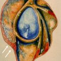
The rotator cuff tendon is also histologically complex. Clark and Harryman described the 5 layers of the rotator cuff. The 2 layers that form the bursal third of the tendon and the layer forming the articular surface of the cuff contain closely packed, well-organized tendon fibers. The 2 layers in the center of the cuff contain less-organized fibers mixed with connective tissue. This anatomic feature is important when looking at MR images of the rotator cuff because it explains why the 2 surfaces of the cuff are low signal, whereas the central portion is intermediate signal on T2-weighted images ( Fig. 2 ).
The various histologic layers of the rotator cuff also result partly from other ligaments combining with the lateral portion of the supraspinatus tendon to form the cuff. For example, the collagen fibers from the joint capsule merge with the undersurface of the supraspinatus tendon to contribute to layer 5. Another important contributor to the rotator cuff is the coracohumeral ligament, which extends posteriorly and divides to send fibers above and below the supraspinatus tendon, thereafter merging with the tendon to form the superficial layer 1 and the deep layer 4 of the rotator cuff. The fibers in layer 4 can form a 1-cm-wide, focally thickened band extending from front to back across the supraspinatus and infraspinatus portions of the cuff, called the ligamentum semicirculare humeri or the rotator cable. The rotator cable can sometimes be seen on MR images as a low-signal band within the cuff, close to the articular surface, running perpendicular to the long axis of the supraspinatus and infraspinatus tendons ( Fig. 3 ).
MR imaging of the rotator cuff
MR imaging of the shoulder is usually performed with the patient supine and the humerus comfortably externally rotated. Slight external rotation aligns the supraspinatus tendon with the blade of the scapula so that it can be optimally evaluated with standard oblique coronal and oblique sagittal planes. If the humerus is allowed to be in internal rotation, the lateral tendon curves out of the oblique coronal plane and causes partial averaging of the tendon with muscle, reducing the accuracy for diagnosing cuff tears.
Although there is no single preferred protocol for imaging the rotator cuff, there are several basic principles. The supraspinatus tendon is the main structure involved in patients with rotator cuff disease and external impingement, so the coronal and sagittal imaging planes should be oriented relative to the long axis of the supraspinatus muscle. High accuracy for diagnosing rotator cuff tears has been reported with fat-suppressed, fast spin-echo, T2-weighted sequences in the oblique, coronal, and oblique sagittal planes. A non–fat-suppressed, short echo time (TE) sequence should also be obtained to evaluate for muscle fatty atrophy. There is more variability in the preferred axial pulse sequence, and the selection of the type of sequence usually depends on reader preference and machine capability.
Normal Rotator Cuff Anatomy and MR Appearance
The rotator cuff is a fairly complex structure composed of 4 tendons: the supraspinatus, the infraspinatus, the teres minor, and the subscapularis tendons. Most infraspinatus tears and many subscapularis tendon tears occur adjacent to supraspinatus tendon tears as part of massive rotator cuff tears. Teres minor tendon tears are rare, although denervation atrophy of the teres minor is not uncommon.
The supraspinatus muscle originates from the posterior upper scapula and extends laterally above the scapular spine to insert onto the superior facet of the greater tuberosity of the humerus. The lateral supraspinatus tendon merges with the more posterior infraspinatus tendon in the lateral 1.5 cm prior to insertion, and this forms a tendinous cuff. The supraspinatus has 2 different tendons laterally. The main tendon forms medially within the mid-substance of the muscle but then lies progressively more anteriorly within the muscle as it moves laterally until it forms the anterior half of the supraspinatus portion of the rotator cuff. The more posterior part of the muscle forms a much shorter aponeurosis that is only about 2 cm in length. It merges laterally with the main anterior supraspinatus tendon anteriorly, and with the infraspinatus tendon posteriorly ( Fig. 1 ).
The rotator cuff tendon is also histologically complex. Clark and Harryman described the 5 layers of the rotator cuff. The 2 layers that form the bursal third of the tendon and the layer forming the articular surface of the cuff contain closely packed, well-organized tendon fibers. The 2 layers in the center of the cuff contain less-organized fibers mixed with connective tissue. This anatomic feature is important when looking at MR images of the rotator cuff because it explains why the 2 surfaces of the cuff are low signal, whereas the central portion is intermediate signal on T2-weighted images ( Fig. 2 ).
The various histologic layers of the rotator cuff also result partly from other ligaments combining with the lateral portion of the supraspinatus tendon to form the cuff. For example, the collagen fibers from the joint capsule merge with the undersurface of the supraspinatus tendon to contribute to layer 5. Another important contributor to the rotator cuff is the coracohumeral ligament, which extends posteriorly and divides to send fibers above and below the supraspinatus tendon, thereafter merging with the tendon to form the superficial layer 1 and the deep layer 4 of the rotator cuff. The fibers in layer 4 can form a 1-cm-wide, focally thickened band extending from front to back across the supraspinatus and infraspinatus portions of the cuff, called the ligamentum semicirculare humeri or the rotator cable. The rotator cable can sometimes be seen on MR images as a low-signal band within the cuff, close to the articular surface, running perpendicular to the long axis of the supraspinatus and infraspinatus tendons ( Fig. 3 ).
MR imaging of external impingement
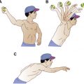
External impingement is generally considered to be a clinical diagnosis. The condition is defined as a chronic ache in the lateral aspect of the shoulder aggravated by elevation of the arm. During forward flexion and abduction, patients usually complain of an arc between 60° and 120° in which the pain is most severe. The pain may also be severe at night and can awaken the patient from sleep.
There are 2 physical examination tests used to confirm external impingement. The Neer impingement test involves forward flexion of the arm with the elbow extended to 180°, with pain elicited at maximal elevation. The Hawkins-Jobe test begins with the shoulder flexed forward at 90° and the elbow bent, with pain elicited with internal rotation, abduction, and increased forward flexion. External impingement is further confirmed if there is decreased pain after a lidocaine injection into the SASD bursa, the so-called impingement test.
The MR imaging findings of a patient with external impingement are variable, and can range from normal to tendinopathy to a rotator cuff tear. There are several MR image findings that are useful to note even in patients with no indications of rotator cuff disease. One of these is abnormal fluid in the SASD bursa, which can indicate SASD bursitis and is often present in patients with symptomatic impingement syndrome. Although a small amount of fluid within the bursa is common, an effusion defined by a width greater than 3 mm suggests bursitis. Other findings associated with SASD bursitis include fluid medial to the acromioclavicular (AC) joint or in the anterior portion of the bursa. Another finding that has been associated with impingement pain is a decreased (≤7 mm) acromiohumeral distance in the absence of a rotator cuff tear.
The most controversial MR imaging findings of impingement involve coracoacromial arch structures. Most investigators agree that resecting large AC joint inferior osteophytes and a type 3, downsloping, anterior acromion process, can slow the progression of rotator cuff disease or improve the healing of a surgically repaired tear. It is less clear whether abnormalities of the coracoacromial arch by themselves cause external impingement syndrome, or if impingement can be diagnosed on an MR image simply by identifying these abnormalities. The following paragraphs describe the MR imaging appearance and summarize the literature on the main coracoacromial arch abnormalities: AC joint osteophytes, anterior hooked acromion process, lateral downsloping of the acromion, os acromiale, and a thickened coracoacromial ligament.
Several investigators have reported that inferiorly directed AC joint osteophytes (especially those ≥3 mm) are associated with impingement and rotator cuff tears ( Fig. 4 ). Other investigators, however, found that AC joint osteoarthritis is very common in patients who are either asymptomatic or have only AC joint tenderness, and have noted that resection of the AC joint leads to instability of the joint and poor outcomes. With newer capsule-sparing and coplaning techniques, symptomatic instability of the distal clavicle after AC joint decompression surgery has been reduced. Most investigators believe it is useful to note the inferiorly directed osteophytes of the AC joint that are larger than 3 mm and indent the rotator cuff on the MR image, because most surgeons resect them at surgery in patients with external impingement syndrome.
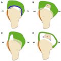
Many articles have been written on the hooked or type 3 anterior acromion process ( Fig. 5 ). Charles Neer popularized the theory that the anterior acromion process was the major extrinsic cause of external impingement and rotator cuff disease. Bigliani and colleagues later proposed a classification system for the anterior acromion process as seen on the coracoacromial arch radiographic view, whereby a downward-hooked acromion process was classified as type 3 and was thought to be most associated with rotator cuff tears. Several other investigators have also found an association between a hooked anterior acromion process and rotator cuff tears. Some also believe that there is a similar association between a downward-tilted anterior acromion process and rotator cuff tears.
There are many articles, however, that dispute the concept that a hooked anterior acromion process causes subacromial impingement syndrome or rotator cuff tears. Several articles have argued that many cases of hooked acromion are actually enthesophytes at the origin of the coracoacromial ligament. These enthesophytes create an extended, more rigid, anterior lip of the acromion process but do not by themselves indent the supraspinatus tendon. The prevalence of the type 3 acromion process has also been shown to increase with age. The association with rotator cuff tears could have occurred because cuff tears are also more common with increasing age or because these enthesophytes are more likely to develop in patients with rotator cuff tears. Several investigators have observed high interobserver variability in classifying the anterior acromion and have noted that slight differences in radiographic technique can alter the appearance and classification assigned to an acromion process.
Matsen and colleagues consider that a downturned, anterior acromion may play a role in impingement pain but only secondarily, with the primary contribution coming from a tight posterior capsule or weak humeral depressors. These investigators propose that a tight posterior capsule or weak humeral head depressors result in a slight anterosuperior shift of the humeral head with arm elevation. Because the humeral head does not remain concentric with the undersurface of the acromion process during arm elevation, the rotator cuff tendon now rubs against and is compressed and abraded by the anterior acromion process, leading to impingement pain and rotator cuff tears.
Whatever the explanation, when treating patients with a rotator cuff tear or external impingement, most surgeons remove the enthesophyte and perform debridement of the downward-curved anterior acromial undersurface to flatten it. This anterior subacromial decompression procedure has been shown to improve impingement pain, even in patients with congenital subacromial stenosis.
Lateral downsloping of the lateral acromion process has also been described by several investigators as a potential cause of impingement pain. Other reports have found that a large lateral extension of the acromion process, or a lateral heel-type spur, can be associated with rotator cuff tears. Prior to Neer’s 1972 article, many impingement patients had lateral acromion resection but did not achieve much pain relief. Neer showed that impingement pain arises mainly from forward flexion of the arm and not from abduction; he therefore disputed the notion that the lateral aspect of the acromion plays a major role in impingement pain for most patients. Other investigators have confirmed that a lateral downsloping acromion does not correlate with impingement pain or rotator cuff tears. Most surgeons are reluctant to resect the lateral portion of the acromion process, except in cases where there is severe lateral downsloping, clearly compressing and potentially abrading the rotator cuff.
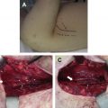
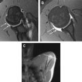
An unfused anterior acromion process or os acromiale has also been associated with impingement syndrome and rotator cuff tears. The theory is that the deltoid muscle attached to a mobile os acromiale contracts and pulls down the os, causing it to bang against the SASD bursa and the rotator cuff to create impingement pain and tears. Some investigators, however, have found no association between os acromiale and rotator cuff tears. Even if there is no association, it is important to identify an os acromiale on the MR image especially if there is a pseudarthrosis with fluid signal at the nonunion, because surgeons need to decide whether to resect, fuse, or leave alone the os at the time of arthroscopy ( Fig. 6 ).
MR imaging of rotator cuff tendinopathy and intratendinous tears
Rotator cuff tendinopathy is usually seen in patients older than 40 years, with a reported average age of about 50 years. There are 2 hallmarks of rotator cuff tendinopathy on MR images: (1) abnormal increased signal within the substance of the cuff without extension to either the articular or bursal surface, and (2) swelling or increased thickness of the tendon ( Fig. 7 ). The increased signal within the cuff has been shown to be caused by mucoid and eosinophilic degeneration, splits in the tendon fibers, increased type III collagen, and degradation of mucopolysaccharide with increased glycosaminoglycan and proteoglycan. Tendinopathy findings on MR images do not necessarily correlate with pain, but the changes are often seen in patients with impingement syndrome.
Related posts:
 Internal Impingement Syndromes
 The Throwing Shoulder: the Orthopedist Perspective
 The Rotator Cable: Magnetic Resonance Evaluation and Clinical Correlation
 Magnetic Resonance Imaging of the Pediatric Shoulder
 Entrapment Neuropathies of the Shoulder
 Anatomic Variants and Pitfalls of the Labrum, Glenoid Cartilage, and Glenohumeral Ligaments
Stay updated, free articles. Join our Telegram channel

Full access? Get Clinical Tree


