The rotator cable is an extension of the coracohumeral ligament coursing along the undersurface of the supraspinatus and infraspinatus tendons. The rotator cable is thought to play a role in the biomechanical function of the intact and torn rotator cuff. It can be seen on all the imaging planes used for the conventional magnetic resonance imaging of the shoulder. Clinically, the integrity of the rotator cable can play a role in the treatment selection for patients with a rotator cuff tear.
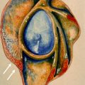
The rotator cable was first described in a study by Clark and Harryman as a fibrous band coursing along the undersurface of the supraspinatus and infraspinatus tendons perpendicular to their fibers and continuous with the coracohumeral ligament anteriorly. Burkhart and colleagues confirmed the presence of this structure and named it the cable, a descriptive term related to its biomechanical role in these investigators’ model of rotator cuff function and failure. The rotator cuff tendon fibers extending distal to the lateral margin of the cable to the greater tuberosity attachment were named the crescent ( Fig. 1 ).

Based on Burkhart’s biomechanical studies, 2 different types of functioning rotator cuff tendons have been described: cable-dominant and crescent-dominant. The cable-dominant rotator cuff was theorized to occur in older persons whose cable absorbs the stress produced by the supraspinatus and infraspinatus tendons while shielding the crescent fibers. The crescent would undergo atrophy and thinning related to the shielding, while assuming a markedly reduced role in the biomechanical function of the rotator cuff. Alternatively, the cable would undergo hypertrophy as it assumed the major role in biomechanical functioning. A crescent-dominant rotator cuff was theorized to occur in younger patients. In this scenario, there was no stress shielding of the crescent by the cable and no associated cable hypertrophy. Thus, the cable would not play a major role in the biomechanical function of the rotator cuff.
The rotator cable also plays an important part in Burkhart’s model of a rotator cuff tear, in which it functions as the loaded cable of a suspension bridge ( Fig. 2 ). In this model, the cable absorbs the compressive and tensile stress produced by the supraspinatus and infraspinatus tendons. The compressive stress is transmitted to its anterior and posterior osseous insertions that serve as the supporting towers where the stress is dissipated. The tensile stress is absorbed and dissipated by the cable itself. According to this model, stress is transferred from the cuff muscles to the rotator cable as a distributed load, thereby stress-shielding the thinner, avascular crescent tissue, particularly in older persons. The cable and its osseous insertions also serve as medial to lateral and anterior to posterior barriers in this model, limiting the propagation of tears involving the crescent while preserving the rotator cuff function.

Anatomy
Gross/Histology
There have been several studies describing the gross and magnetic resonance (MR) anatomy of the rotator cable. Gross studies have shown a close anatomic relationship between the rotator cable and the coracohumeral ligament. The coracohumeral ligament arises from the rotator interval and envelops the rotator cuff with superficial and deep limbs ( Fig. 3 ). The superficial limb is diminutive and lies along the bursal surface of the tendons. The deep limb is thought to represent the cable and tends to be a larger, thicker structure. The anterior insertion site of the cable is found at the greater tuberosity along the anterior margin of the supraspinatus tendon, just posterior to the biceps tendon. The cable then extends posteriorly perpendicular to the long axis of the supraspinatus and infraspinatus tendon fibers, interposed between the rotator cuff undersurface and the joint capsule. The posterior margin of the cable inserts along the inferior border of the infraspinatus tendon. The cable forms the medial margin of a crescent-shaped area that includes the distal fibers of the supraspinatus and infraspinatus, known as the crescent, located approximately 1.1 to 1.5 cm from the greater tuberosity. Studies have shown variable degrees of thickness and width of the cable ranging between 1.2 to 4.7 mm and 4.5 to 12.1 mm, respectively. The crescent includes the critical zone, a hypovascular region of the rotator cuff that tends to undergo attritional change and degenerative tearing over time. Histologic examination has demonstrated the cable as a fibrillar collagenous structure separate from the supraspinatus and infraspinatus tendon fibers.

Imaging
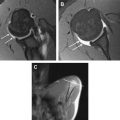
Several studies have examined the imaging appearance of the rotator cuff cable. The rotator cuff cable is consistently seen on an ultrasonogram as a fibrillar structure coursing perpendicular to the supraspinatus and infraspinatus tendons. The rotator cuff cable can be seen in all the main imaging planes used in MR imaging. In both the coronal oblique and the abducted, externally rotated (ABER) planes, the rotator cable appears as a region of hypointense signal intensity along the undersurface of the supraspinatus and infraspinatus tendons that is continuous with the coracohumeral ligament ( Fig. 4 ). The oblique coronal plane provides a cross-sectional view of the rotator cable, and therefore is most useful in the assessment of its craniocaudal thickness and width. In this plane, the cable is typically seen as a rounded focus of hypointense signal on all pulse sequences varying in craniocaudal size from 1 to 5 mm ( Figs. 5 and 6 A). In some instances, however, the cable appears as a broad, dotted line (see Fig. 6 B). In the authors’ experience, a prominent cable is more often visualized among individuals in the fifth to seventh decades of life, who undergo MR imaging of the shoulder in a search for rotator cuff disease. In these cases, the presence of an undersurface fraying and a shallow tearing of the rotator cuff may help highlight the margins of the cable, increasing its conspicuity. Differentiating the rotator cable from the retracted lateral edge of an articular surface partial tear of the supraspinatus tendon can be challenging, and the authors find triangulating the suspected cable in the axial plane most useful. A true cable will be seen extending from its anterior attachment in the greater tuberosity to its posterior oblique facet insertion in the axial images as opposed to the more focal changes seen in a retracted tear of the supraspinatus articular surface. In young adults, the cable may not be as easily discriminated from the adjacent rotator cuff tendon fibers. Inconsistent visualization of the cable in the setting of partial and full-thickness tears of the rotator cuff has been reported.

Related posts:
 Magnetic Resonance Imaging of Rotator Cuff Disease and External Impingement
 Internal Impingement Syndromes
 The Throwing Shoulder: the Orthopedist Perspective
Magnetic Resonance Imaging of the Pediatric Shoulder
The Throwing Shoulder: the Orthopedist Perspective
Magnetic Resonance Imaging of the Pediatric Shoulder
 Entrapment Neuropathies of the Shoulder
 Anatomic Variants and Pitfalls of the Labrum, Glenoid Cartilage, and Glenohumeral Ligaments
Stay updated, free articles. Join our Telegram channel

Full access? Get Clinical Tree


