Both benign and malignant pelvic masses are encountered in the pediatric population. Although ultrasonography remains the modality of choice for initial evaluation of a pediatric pelvic mass, in selected cases magnetic resonance (MR) imaging can add important diagnostic information. MR imaging has several advantages over ultrasonography and computed tomography, including superior contrast resolution and an ability to characterize abnormalities based on unique tissue characteristics. MR evaluation assists in lesion characterization, presurgical planning, and staging when a malignancy is suspected. MR imaging also offers a nonionizing imaging modality for long-term follow-up of patients undergoing therapy for malignant pelvic masses.
Key points
- •
Magnetic resonance (MR) evaluation of pediatric pelvic masses is advantageous, given its excellent soft-tissue contrast resolution and lack of ionizing radiation.
- •
Rhabdomyosarcoma is the most common primary malignant neoplasm of the lower genitourinary tract. There is a peak incidence between 2 and 4 years of life, with a second peak in adolescence.
- •
Most pediatric ovarian masses are benign and functional. Functional ovarian cysts are the most commonly encountered entity and the most common abdominal mass in a newborn female. Malignant ovarian neoplasms are uncommon.
- •
Germ-cell tumors are the most common type of ovarian neoplasm encountered in the pediatric population. The most common type of pediatric ovarian germ-cell tumor is a mature teratoma.
- •
Uterine tumors are uncommon in pediatric patients, but when encountered in this population they are more likely to be malignant.
- •
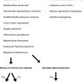
Other miscellaneous tumors found in the pelvis in the pediatric population include those in the presacral space; these can be classified as congenital, developmental, neurogenic, inflammatory, mesenchymal, and osseous (extension of sacral neoplasms).
Introduction
Both benign and malignant pelvic masses are encountered in the pediatric population. Although ultrasonography remains the modality of choice for initial evaluation of a pediatric pelvic mass, magnetic resonance (MR) imaging is often used for further evaluation and lesion characterization. MR evaluation of pediatric pelvic masses is advantageous, given its multiplanar capabilities, excellent soft-tissue contrast resolution, and ability to specifically characterize certain types of tissues (ie, fat and blood). Contrast-enhanced MR imaging assists in mass characterization, presurgical planning, and staging when a malignancy is suspected. The lack of ionizing radiation is another advantage of MR imaging, particularly given the radiosensitivity of the gonads, and when multiple consecutive studies are needed to monitor therapy response in malignant conditions.
Pediatric pelvis masses can arise from a variety of pelvic structures, including the ovaries, lower genitourinary tract, musculature, neurovascular structures, lymph nodes, and adjacent osseous structures. Although the MR appearance of many pelvic masses is nonspecific, some masses demonstrate characteristic imaging features. For example, ovarian teratomas typically demonstrate areas of hyperintense signal on T1-weighted and T2-weighted sequences compatible with fat. Dysgerminomas contain internal fibrovascular septations that enhance on postcontrast sequences. Given its excellent soft-tissue contrast resolution, MR is useful in evaluating the full extent of a mass, including the relationship of the mass with adjacent critical pelvic structures, which may be important for presurgical planning and staging especially when a pelvic malignancy is suspected.
Protocol
Pelvic Mass MR Imaging Protocol
The authors perform MR imaging for pelvic masses at both 1.5 and 3 T, but prefer 3 T imaging because of its improved signal to noise ratio (SNR), allowing for shorter image acquisition times and improved spatial resolution. During imaging, a multichannel torso surface coil is placed over the patient’s pelvis and lower abdomen anteriorly. Parallel imaging can be used to reduce scan times. A routine pelvic mass protocol includes coronal T1-weighted fast spin-echo, coronal short-tau inversion recovery (STIR), axial T2-weighted fat-suppressed fast spin-echo, axial T1-weighted dual-gradient echo in-phase and opposed-phase, axial diffusion-weighted, axial 3D T1-weighted fat-suppressed spoiled gradient recalled echo before intravenous contrast, and axial, sagittal, and coronal postcontrast 3D T1-weighted fat-suppressed spoiled gradient recalled echo acquisitions. In female patients, if a detailed evaluation of the uterus is indicated, the authors include T2-weighted fast spin-echo sequences in short and long axis relative to the uterus, and sagittal T2-weighted fast spin-echo sequence. Details of their institutional pelvic mass protocol are included in Table 1 . Standard intravenous gadolinium-based contrast material dosing is calculated based on the patient’s weight. The authors do not routinely administer glucagon or other antiperistaltic agents before pelvic MR imaging.
| Sequence | Plane | Fat Saturation (Yes/No) | Flip Angle (°) | TE (ms) | TR (ms) | Slice Thickness/Gap (mm) | NSA |
|---|---|---|---|---|---|---|---|
| T1-weighted fast spin-echo | Coronal | No | 90 | 8 | Shortest | 5/1 | 2 |
| Short-tau inversion recovery (STIR) | Coronal | None | 60 | Shortest | 5/1 | 1 | |
| T2-weighted fast spin-echo | Axial | Yes | 90 | 75 | Shortest | 5/1 | 1 |
| T1-weighted dual gradient echo in-phase and opposed-phase | Axial | No | 55 | In-phase = 2.3 Opposed-phase = 1.15 | 180 | 5/0.5 | 1 |
| T2-weighted fast spin-echo a | Sagittal | No | 90 | 75 | Shortest | 5/1 | 1 |
| T2-weighted fast spin-echo a | Short and long axis relative to the uterus | No | 90 | 90 | Shortest | 4/0 | 1 |
| Diffusion-weighted | Axial | Yes | 90 | Shortest | Shortest | 5/0 | 4 |
| Precontrast 3D-SPGR | Axial | Yes | 10 | Shortest | Shortest | 1.5–3 | 1 |
| Postcontrast 3D-SPGR | Coronal, axial, sagittal | Yes | 10 | Shortest | Shortest | 1.5–3 | 1 |
When evaluating bladder masses, dynamic T1-weighted 3D-SPGR postcontrast sequences are performed to evaluate bladder-wall enhancement and tumor extension before excreted contrast reaches the bladder. Dynamic T1-weighted postcontrast sequences in the arterial, venous, and delayed phases are also used to evaluate patients with suspected vascular malformations involving the pelvis.
Ovary
The majority of pediatric ovarian masses are functional and benign. The functional ovarian cyst is the most commonly encountered entity and the most common abdominal mass in a newborn female. Malignant ovarian neoplasms are uncommon, accounting for approximately 1% to 2% of malignant pediatric neoplasms and 10% of all ovarian neoplasms. Ovarian malignancies rarely develop in infants younger than 1 year. Ovarian malignancy is more frequently encountered in older children (age 9–19 years). Suspicion of an ovarian malignancy must be maintained if a girl presents with an abdominopelvic mass, especially in prepubertal girls (between infancy and the onset of puberty), who uncommonly have functional ovarian lesions.
Classification of ovarian tumors is based on their cell of origin and includes germ-cell, epithelial, and sex-cord stromal tumors. Germ-cell tumors are the most common and comprise 60% to 90% of pediatric ovarian neoplasms, followed by sex-cord stromal (10%–13%) and epithelial tumors (5%–11%). Ovarian neoplasms can be either benign or malignant, and often contain cystic and solid components; in general, the solid component is a predictor of malignancy. The most common benign tumors are teratomas, followed by cystadenomas.
A suspected pelvic mass in a female child should be initially investigated with ultrasonography. Depending on the sonographic findings, cross-sectional imaging may be indicated. MR imaging has proved useful in further characterization of ovarian masses, provides staging information, and assists in preoperative planning. Clinical presentations of pelvic masses vary and include nonspecific abdominal pain, increasing abdominal circumference, ovarian torsion, and, occasionally, symptoms related to excess hormone production by the tumor. Treatment is variable depending on the tumor; general treatment options include surgery and chemotherapy. Some malignant tumors such as dysgerminomas and granulosa cell tumors are also radiosensitive. Table 2 summarizes the imaging characteristics of the most common ovarian lesions.
| Ovarian Lesion | Imaging Characteristics |
|---|---|
| Simple cyst | Low signal on T1-weighted and high on T2-weighted sequences with a thin rim of peripheral enhancement |
| Hemorrhagic cyst | Commonly high signal on T1-weighted sequences given presence of blood products, although variable depending on evolution of blood products. Peripheral enhancement common, but no nodular/central enhancement should be present |
| Torsion | Intermediate to high signal on T2-weighted sequences because of edema. Variable degrees of nonenhancement depending on degree of infarction |
| Teratoma | High signal on T1-weighted and T2-weighted sequences given presence of fat. Most present as unilocular cystic masses |
| Dysgerminoma | Solid mass with calcifications and enhancement of fibrous septa |
| Sertoli-Leydig neoplasm | Can have multiple cystic areas and demonstrate areas of low signal on T1-weighted and T2-weighted sequences, owing to fibrosis |
| Serous epithelial | Unilocular or multilocular components with thin septations; usually large at presentation. Papillary and solid components more predictive of malignancy |
| Mucinous epithelial | Typically multilocular with the multiple cysts containing fluid demonstrating variable signal; usually large at presentation. Papillary and solid components more predictive of malignancy |
Functional Ovarian Lesions
Functional ovarian lesions such as follicular cysts and corpus luteal cysts are the most common adnexal masses seen in children, accounting for approximately 45% of all adnexal abnormalities. Functional ovarian lesions are occasionally encountered in the neonatal period when there is exposure to maternal hormones, but are most common in the postmenarchal period. The functional ovarian cyst is also the most commonly encountered intra-abdominal mass in females in utero. Most cysts encountered are simple, but complex cysts also occur. Any cystic lesion identified between infancy and the onset of puberty should be viewed with a higher degree of suspicion, as functional ovarian cysts are only rarely encountered in this age group.
Although ultrasonography is typically adequate for detection and characterization of ovarian cysts, MR imaging can be useful in certain cases. MR imaging can confirm the presence of a cyst when the ultrasound imaging characteristics are atypical or when an underlying mass is suspected. A simple ovarian cyst should demonstrate homogeneous T1-weighted hypointense and T2-weighted hyperintense signal characteristics with uniform peripheral or rim enhancement after the administration of gadolinium-containing contrast material ( Fig. 1 ). Functional hemorrhagic cysts classically have been described to demonstrate hyperintense signal on T1-weighted images ; however, the MR appearance of hemorrhagic cysts can vary somewhat depending on the evolutionary stage of blood products. Kanso and colleagues described only 36% of hemorrhagic cysts demonstrating intermediate or hyperintense signal on T1-weighted sequences ( Fig. 2 ). A corpus luteum cyst that has involuted can have wavy and irregular borders and may also demonstrate heterogeneous signal, depending on the presence of blood products (see Fig. 2 ). There should be no central or nodular enhancement in any of these benign, functional ovarian cysts.
Treatment is usually conservative unless there is associated ovarian torsion or the cyst reaches a particular size that would significantly increase the risk of torsion.
Ovarian Torsion
Ovarian torsion is a surgical emergency that results from twisting of the ovary on its vascular pedicle with resultant vascular compromise. Fifteen percent of ovarian torsion occurs in the pediatric population. Ovarian torsion is commonly associated with other ovarian abnormalities in the adult population; however, in children ovarian torsion is more commonly associated with benign lesions or normal ovaries. Ovarian torsion can occur from inherent hypermobility of the ovary, or secondary to a mass acting as a lead point. Clinical presentations include abdominal pain, nausea and vomiting, and palpable mass (detected in 40%–70% of patients).
Ultrasonography is the initial modality of choice in suspected ovarian torsion, and is usually the only imaging test obtained before surgical intervention. In rare cases, MR imaging with contrast may assist in the diagnosis when sonographic findings are confounding or when there is suspicion for an underlying mass acting as a lead point. With torsion, ovarian follicles may migrate to the periphery of the ovary, which has been described as resembling a “string of pearls.” Postcontrast imaging will assist with detection of solid/nodular components if an underlying mass is present. Nonenhancement of ovarian parenchyma is compatible with infarction ( Fig. 3 ). The torsed ovary can have variable degrees of T2-weighted signal intensity from intermediate to high, depending on the amount of edema present. Torsion can occur in utero. After birth, the nonviable ovary can appear as a complex cystic lesion and may eventually involute into a calcified mass.
The treatment for ovarian torsion is emergent surgery. Early diagnosis is critical for preserving fertility and maximizing chances of ovarian salvage. Unfortunately, one large series reported that approximately 58% of patients are treated with oophorectomy because of delays in diagnosis.
Germ-Cell Neoplasms
Germ-cell tumors are the most common type of ovarian neoplasm encountered in the pediatric population, in contrast to the adult population in which the majority of ovarian malignancies are epithelial in origin. Germ-cell tumors comprise between 60% and 90% of pediatric ovarian malignancies, depending on the cited study. Children with gonadal dysgenesis are at increased risk for developing germ-cell tumors.
Germ-cell tumors include mature teratomas, immature teratomas, dysgerminomas, endodermal sinus tumors, embryonal carcinomas, and choriocarcinomas. The most common type of pediatric ovarian germ-cell tumor is a mature teratoma, which is benign. All of the other types are malignant and have the potential to metastasize to the liver, lungs, and peritoneum.
Mature teratomas account for 67% of pediatric ovarian tumors and are bilateral 25% of the time. They comprise at least 2 of the 3 embryonic germ-cell layers: ectoderm, mesoderm, and endoderm. Imaging features of mature teratomas range from completely cystic lesions to solid, predominantly fat-containing masses. Most teratomas present as a unilocular cystic mass ( Fig. 4 ) and commonly contain a Rokitansky nodule (a soft-tissue plug containing hair, fat, and calcific and/or sebaceous components), which extends into the lumen of the cyst ( Fig. 4 ). The imaging appearance of a mature teratoma varies, particularly on ultrasonography, depending on the size of the Rokitansky nodule and the distribution of calcifications and fat. However, their appearance on MR imaging is more characteristic. The presence of macroscopic fat in an ovarian tumor is diagnostic of a mature teratoma, and can be exquisitely demonstrated with the use of in-phase/opposed-phase imaging and fat-suppressed MR sequences ( Fig. 5 ). The fatty component of the tumor will demonstrate hyperintense signal on T1-weighted and T2-weighted non–fat-suppressed sequences, and will subsequently lose signal with fat suppression. Lesions that contain hemorrhage or proteinaceous material will demonstrate hyperintense signal on T1-weighted sequences both with and without fat suppression.
The remaining tumors in this category, including dysgerminomas, yolk sac tumors, and choriocarcinomas, generally demonstrate a nonspecific imaging appearance. However, MR imaging is useful for preoperative planning and staging. Dysgerminoma is the most common malignant ovarian neoplasm ( Fig. 6 ). Imaging may demonstrate a solid mass with small calcifications. Dysgerminomas demonstrate low signal intensity on T1-weighted sequences and intermediate to high signal on T2-weighted sequences, depending on the amount of necrosis present. Intralesional fibrous septa demonstrate low signal intensity on T2-weighted sequences and enhance on postcontrast sequences. Other types of germ-cell tumors may be distinguished by their hormonal markers. For example, endodermal sinus tumors may have elevated levels of α-fetoprotein (AFP), whereas embryonal carcinomas may have elevated levels of both AFP and β–human chorionic gonadotropin.
Stromal Cell Tumors
Tumors of stromal cell origin constitute the second most common category of pediatric ovarian neoplasms. Although there are several types, the most commonly encountered are granulosa cell tumors (75%) followed by Sertoli-Leydig tumors (25%). Because of their functional nature, patients with granulosa tumors can present with precocious puberty arising from the estrogen exposure. On the other hand, oligomenorrhea and virilization can be presenting features in patients with Sertoli-Leydig tumors, as a result of excess androgen production. Granulosa tumors have a variable MR appearance, and can present as cystic or solid masses containing areas of hemorrhage and infarction. Sertoli-Leydig tumors are well defined, can contain multiple cystic areas, and can demonstrate hypointense signal on T1-weighted and T2-weighted sequences owing to fibrosis.
Epithelial Tumors
Epithelial ovarian neoplasms are the least common group of pediatric ovarian malignancies. The majority of epithelial tumors are benign (approximately 58%) followed by those categorized as having borderline to low malignant potential. High-grade malignant epithelial tumors are rare. Serous and mucinous cystadenomas, the most common types of epithelial neoplasms, are typically large at presentation and can present with ovarian torsion. Serous cystadenomas usually have a significant unilocular or multilocular cystic component and thin septations. Mucinous cystadenomas are typically multilocular, septated masses, with the multiple cysts containing fluid demonstrating varying signal intensities ( Fig. 7 ). Papillary projections and solid, enhancing components may be present and are more predictive of malignancy ( Fig. 8 ).
Related posts:
 Magnetic Resonance Imaging of the Pediatric Liver
 Magnetic Resonance Imaging of the Pediatric Liver
Magnetic Resonance Imaging of the Pediatric Kidney
Magnetic Resonance Imaging of Perianal and Perineal Crohn Disease in Children and Adolescents
Magnetic Resonance Imaging of Anorectal Malformations
Advanced Techniques in Pediatric Abdominopelvic Oncologic Magnetic Resonance Imaging
Magnetic Resonance Imaging of the Pediatric Liver
Magnetic Resonance Imaging of the Pediatric Kidney
Magnetic Resonance Imaging of Perianal and Perineal Crohn Disease in Children and Adolescents
Magnetic Resonance Imaging of Anorectal Malformations
Advanced Techniques in Pediatric Abdominopelvic Oncologic Magnetic Resonance Imaging
Stay updated, free articles. Join our Telegram channel

Full access? Get Clinical Tree


